. 2012 Feb;86(4):1955-70.
doi: 10.1128/JVI.05949-11. Epub 2011 Dec 7.

Multiple layers of CD80/86-dependent costimulatory activity regulate primary, memory, and secondary lymphocytic choriomeningitis virus-specific T cell immunity

Affiliations

Multiple layers of CD80/86-dependent costimulatory activity regulate primary, memory, and secondary lymphocytic choriomeningitis virus-specific T cell immunity

Jens Eberlein et al. J Virol. 2012 Feb.

Abstract

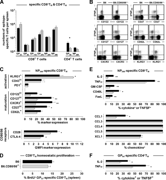
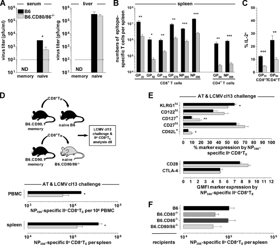
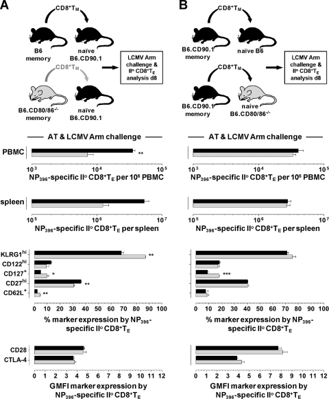
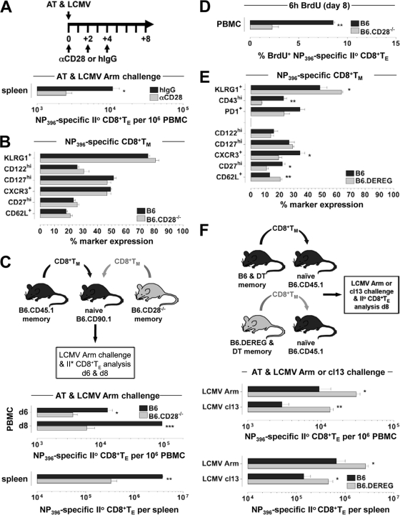
The lymphocytic choriomeningitis virus (LCMV) system constitutes one of the most widely used models for the study of infectious disease and the regulation of virus-specific T cell immunity. However, with respect to the activity of costimulatory and associated regulatory pathways, LCMV-specific T cell responses have long been regarded as relatively independent and thus distinct from the regulation of T cell immunity directed against many other viral pathogens. Here, we have reevaluated the contribution of CD28-CD80/86 costimulation in the LCMV system by use of CD80/86-deficient mice, and our results demonstrate that a disruption of CD28-CD80/86 signaling compromises the magnitude, phenotype, and/or functionality of LCMV-specific CD8(+) and/or CD4(+) T cell populations in all stages of the T cell response. Notably, a profound inhibition of secondary T cell immunity in LCMV-immune CD80/86-deficient mice emerged as a composite of both defective memory T cell development and a specific requirement for CD80 but not CD86 in the recall response, while a related experimental scenario of CD28-dependent yet CD80/86-independent secondary CD8(+) T cell immunity suggests the existence of a CD28 ligand other than CD80/86. Furthermore, we provide evidence that regulatory T cells (T(REG)s), the homeostasis of which is altered in CD80/86(-/-) mice, contribute to restrained LCMV-specific CD8(+) T cell responses in the presence of CD80/86. Our observations can therefore provide a more coherent perspective on CD28-CD80/86 costimulation in antiviral T cell immunity that positions the LCMV system within a shared context of multiple defects that virus-specific T cells acquire in the absence of CD28-CD80/86 costimulation.


Figures

Fig 1
Fig 1
CD80/86 expression by major immune cell subsets. Spleen cells from naïve B6 and B6.CD80/86−/− mice were stained for various surface markers as well as CD80 or CD86. (A) Histograms were gated on the indicated immune cell subsets by employing the following gating strategies: CD8+ T cells, CD3ϵ+ CD8+; CD4+ T cells, CD3ϵ+ CD4+; B cells, CD19+; NK cells, CD3ϵ NK1.1+; CD11b+ myeloid cells, CD3ϵ NK1.1 CD11b+ (containing monocytes, macrophages, neutrophils, and DC subsets); DCs, CD3ϵ/CD11chi. Gray histograms, control strains (B6.CD80/86−/−); black tracings, experimental strains (B6). (B) Absolute numbers of CD80- or CD86-expressing cell subsets in the spleens of unmanipulated B6 mice. (C) Distribution of CD80 and CD86 expressions among immune cell subsets in B6 mice. The absolute numbers of CD80- or CD86-expressing T cells, B cells, NK cells, CD11b+ myeloid cells, and DCs were added and set at 100%; the relative contribution of individual immune cell subsets to overall CD80 or CD86 expression was then calculated accordingly, and error bars were omitted for clarity. (D) CD80 and CD86 expressions by naïve (CD44lo) and memory phenotype (CD44hi) T cell subsets. All data are standard errors of the means (SEM) determined for 3 mice/group.
Fig 2
Fig 2
Virus control and effector T cell responses in the absence of CD80/86. (A) B6 mice (black) and B6.CD80/86−/− mice (gray) were infected with 2 × 10⁵ PFU LCMV Armstrong (Arm) i.p., followed by determinations of viral titers in the spleen at the indicated time points. The dotted line indicates the detection threshold of 2 × 10² PFU/mg tissue. (B) Rapid death of both B6 (black) and B6.CD80/86−/− (gray) mice following i.c. infection with 103 PFU LCMV Arm. (C) Frequencies of CD44hi T cells (left) and CD62Lhi or CD127+ T cells within the CD44hi T cell compartments (right) of B6 mice (black) and B6.CD80/86−/− mice (gray) 8 days after LCMV Arm i.p. challenge. ns, not significant. (D) Frequencies (left) and absolute numbers (right) of epitope-specific CD8+ and CD4+ T cells in the spleen determined by intracellular IFN-γ staining. All bar diagrams display SEM (3 to 5 mice/group). (E) Phenotypes of NP396-specific CD8+ TE (plots are gated on CD8+ T cells on day 8 after LCMV challenge). (F) Summary of phenotypic analyses of DbNP396+ CD8+ TE (day 8) in B6 (black) and B6.CD80/86−/− (gray) mice. CTLA-4 expression levels refer to both surface and intracellular CTLA-4 levels, and no significant differences were recorded for CD49d, CD223, or CD244 expression (not shown). (G) Inducible cytokine, TNFSF, and chemokine production assessed by brief in vitro restimulation of CD8+ TE with the NP396 peptide as detailed in Materials and Methods. The bar diagrams display the fraction of NP396-specific (IFN-γ+) CD8+ TE producing the indicated additional cytokines, TNFSFs, or chemokines (representative data from 2 to 3 experiments are SEM for 3 to 4 mice/group).
Fig 3
Fig 3
Role of T cell-expressed CD80/86 and TREGs in generation of primary CD8+ TE responses. (A) To generate p14 chimeras, 2 × 10³ p14 TCRtg CD8+ T cells purified from naïve CD90.1+ p14 mice were adoptively transferred into congenic B6 or B6.CD80/86−/− mice that were subsequently infected with 2 × 10⁵ PFU LCMV Arm i.p. (B and C) Phenotypic (B) and functional (C) analyses of splenic p14 TE were conducted 8 days later (data are SEM [n = 3]). ND, not determined; GMFI, geometric mean of fluorescence intensity. (D and E) Spleen cells obtained from naïve B6 and B6.CD80/86−/− mice were stained for CD4 and CD25 as well as intracellular FoxP3. The representative dot plots are gated on CD4+ cells (D), and the bar diagram (E) shows the absolute numbers of indicated CD4+ T cell subsets in the spleens of these mice (ns, not significant). Note the ∼7-fold reduction of CD4+ CD25+ FoxP3+ TREGs in naïve B6.CD80/86−/− mice (no differences in the level of FoxP3 mean fluorescence intensity [not shown]). (F) B6 and B6.DEREG mice were treated with diphtheria toxin (DT) on days −1, +2, and +5 (1.0 μg DT i.p.) in relation to LCMV infection. Dot plots are gated on CD4+ T cells analyzed right before the first DT injection (day −1) and 1 day after LCMV challenge (day +1). The bar diagram enumerates total and NP396-specific CD8+ T cells in the blood of DT-treated B6 (black) and B6.DEREG (gray) mice 8 days after infection. (G) Phenotypes of NP396-specific CD8+ TE analyzed on day 8 after challenge (peripheral blood mononuclear cells [PBMC]) (all data are SEM [n = 4 to 5]).
Fig 4
Fig 4
Phenotypic and functional alterations of LCMV-specific CD4+ TE generated in the absence of CD80/86. (A) Eight days after LCMV challenge, spleen cells were stained with I-AbGP66 and control (I-AbhCLIP87) tetramers as detailed in Materials and Methods. Plots are gated on B220/CD4+ T cells. Values indicate percentages (SEM) of I-AbGP66+ CD4+ TE; similar results were obtained with I-AbGP61-80 tetramers produced in the laboratory (not shown). (B and C) Phenotypes of GP66-specific CD4+ TE (day 8 after challenge). Dot plots are gated on B220/CD4+ T cells. (D) Functional profiles of GP64-specific CD4+ TE evaluated after 5 h of in vitro peptide stimulation. The bar diagrams depict the fractions of GP64-specific (IFN-γ+) CD4+ TE synthesizing the indicated cytokines, TNFSFs, or chemokines. (E) Mixed peripheral chimeras generated by the combination of B cell-depleted B6.CD45.1 and B6.CD80/86−/− spleen cells at a ratio of 1:1 and AT into OT-I recipients heterozygous at the CD90 locus (CD90.1 × CD90.2). TCRtg OT-I mice, due to their restricted T cell repertoire, cannot generate LCMV-specific CD8+ or CD4+ TE responses such that all LCMV-specific T cells have to be recruited from the donor T cell pool (; Eberlein and Homann, unpublished). Eight days after LCMV challenge, CD4+ donor TE expansions in peripheral blood were quantified (dot plot gated on CD90.1/CD4+ donor CD4+ T cells); similar results were obtained for NP396-specific CD8+ TE, and the chimeric mice controlled the LCMV infection, as assessed by serum plaque assays (not shown). Representative data are SEM (for n ≥ 3 mice/group for all experiments).
Fig 5
Fig 5
Altered phenotypic and functional properties of LCMV-specific TM generated in B6.CD80/86-deficient mice. (A) Absolute numbers of epitope-specific CD8+ and CD4+ TM in the spleens of B6 (black) and B6.CD80/86−/− (gray) mice (84 days postinfection [dpi]) calculated after restimulation with different peptides and intracellular IFN-γ expression analysis. (B and C) Representative dot plots (gated on CD8+ T cells) and summary of DbNP396+ CD8+ TM phenotypes (54 dpi). No differences were observed for CD223 or CD244 expression (not shown). (D) Homeostatic proliferation of GP33-specific CD8+ TM determined after a 7-day BrdU pulse (77 to 84 dpi), as detailed in Materials and Methods. (E) Functional profiles of NP396-specific CD8+ TM analyzed at 54 dpi (cytokines and TNFSFs) or 62 dpi (chemokines). (F) Induced IL-2 and TNF-α production by GP64-specific (IFN-γ+) CD4+ TM determined at 54 dpi. All data are SEM for ≥3 mice/group and independent experiments performed 2 to 3 times.
Fig 6
Fig 6
CD80/86, immune protection, and secondary TE reactivity under conditions of LCMV cl13 rechallenge. (A) Naïve B6 and B6.CD80/86−/− as well as LCMV-immune (“memory”) B6 and B6.CD80/86−/− mice (93 dpi) were challenged with 2 × 10⁶ PFU LCMV clone 13 i.v., and infectious virus titers in serum and liver were determined 6 days later. The dotted line indicates the detection threshold of 2 × 10² PFU/mg tissue. ND, not detected. Data are SEM for 3 to 4 mice/group. (B) Absolute numbers of secondary epitope-specific (IFN-γ+) CD8+ and CD4+ TE in the spleen cl13 rechallenge of LCMV Arm-immune mice (please note that these experiments were conducted 6 days after rechallenge, i.e., 2 days earlier than most of the subsequent analyses, which were performed on day 8). (C) Induced IL-2 production by secondary GP33-specific CD8+ and GP64-specific CD4+ TE. (D) CD8+ T cells were enriched from LCMV-immune wt donors (193 dpi) by combined CD4/B220 depletion, and populations containing 104 DbNP396+ CD8+ TM were transferred into naïve B6 or B6.CD80/86−/− recipients that were subsequently challenged with 2 × 10⁶ PFU LCMV cl13 i.v. Secondary donor CD8+ TE expansions in blood and spleen were quantified 8 days later. (E) Phenotypes of secondary DbNP396+ CD8+ TE. (F) Experimental design as detailed above for panel D, using enriched LCMV-immune wt donor populations containing 2 × 10³ DbNP396+ CD8+ TM (54 dpi) and the indicated naïve B6, B6.CD80−/−, B6.CD86−/−, and B6.CD80/86−/− recipients; cl13 rechallenge; and day 8 analysis (n = 3 mice/group for all AT experiments). II°, secondary.
Fig 7
Fig 7
CD80/86 deficiency and secondary CD8+ TE reactivity under conditions of LCMV Arm rechallenge. (A) CD8+ T cells were enriched from LCMV-immune B6 and B6.CD80/86−/− donors (62 to 64 dpi) by CD4+ T/B cell depletion, and populations containing 104 DbNP396+ CD8+ TM were transferred into congenic wt recipients, followed by LCMV Arm challenge (2 × 10⁵ PFU i.p.) and analyses of secondary CD8+ TE expansions and phenotypes 8 days later (the phenotypes of concurrent primary DbNP396+ CD8+ TE generated by the CD90.1+ hosts were identical in recipients of B6 and B6.CD80/86−/− CD8+ TM [not shown]). (B) Experimental setup similar to that described above for panel A but using only LCMV-immune wt donors (193 dpi) and B6 versus B6.CD80/86−/− recipients. The same results were obtained in four independent experiments, including cotransfer experiments in which CD80/86−/− and congenic (CD90.1) wt CD8+ TM were monitored within the same congenic (CD45.1) hosts (not shown). II°, secondary.
Fig 8
Fig 8
Roles of CD28 and TREGs in regulation of secondary CD8+ TE proliferation and expansion. (A) AT/rechallenge experiments conducted with 2 × 10³ DbNP396+ wt CD8+ TM (137 dpi) and congenic wt recipients in the presence of an anti-CD28 blockade or hamster IgG control treatment. The diagram depicts the schedule of antibody administration in relation to AT/LCMV Arm challenge on day 0 (analyses were performed on day 8). Similar results were obtained in 4 independent experiments (n = 3 mice/group for all AT experiments). (B) Phenotypes of specific CD8+ TM (53 dpi) in LCMV Arm-immune CD28-deficient mice. (C) CD8+ TM (∼340 dpi) were enriched from LCMV-immune B6.CD45.1 and B6.CD28−/− donors and mixed, and populations containing 104 DbNP396+ CD8+ TM each were transferred into naïve B6.CD90.1 congenic recipients prior to LCMV Arm challenge. Quantifications of secondary CD8+ TE expansions were conducted on day 6 and day 8. (D) Proliferation of secondary wt and CD28−/− CD8+ TE among peripheral blood mononuclear cells assessed following a 6-h in vivo BrdU pulse (8 dpi), as detailed in Materials and Methods. Similar results were obtained for CD8+ TE recovered from other tissues and/or specific for other LCMV determinants (not shown). (E) Phenotypes of blood-borne DbNP396+ CD8+ TM (59 dpi) in B6 and B6.DEREG mice treated with DT only during the early primary CD8+ TE response (day −1 to day +5) (Fig. 3F). (F) Splenic CD8+ T cells were enriched (CD4+ T/B cell depletion) from LCMV Arm-immune B6 versus B6.DEREG mice (66 dpi) originally treated with DT as indicated above, and CD8+ T cell populations containing 104 DbNP396+ CD8+ TM each were transferred into naïve CD45.1 congenic recipients that were subsequently challenged with LCMV Arm or cl13 and analyzed 8 days later. Note that the recipient mice maintained intact TREG compartments throughout these AT/rechallenge experiments (data are SEM for 3 mice/group). II°, secondary.

Similar articles

Cited by

References

    1. Andreasen SO, Christensen JE, Marker O, Thomsen AR. 2000. Role of CD40 ligand and CD28 in induction and maintenance of antiviral CD8+ effector T cell responses. J. Immunol. 164:3689–3697 - PubMed
    1. Arens R, et al. 2011. B7-mediated costimulation of CD4 T cells constrains cytomegalovirus persistence. J. Virol. 85:390–396 - PMC - PubMed
    1. Arens R, et al. 2011. Differential B7-CD28 costimulatory requirements for stable and inflationary mouse cytomegalovirus-specific memory CD8 T cell populations. J. Immunol. 186:3874–3881 - PMC - PubMed
    1. Badovinac VP, Haring JS, Harty JT. 2007. Initial T cell receptor transgenic cell precursor frequency dictates critical aspects of the CD8(+) T cell response to infection. Immunity 26:827–841 - PMC - PubMed
    1. Bertram EM, et al. 2004. A switch in costimulation from CD28 to 4-1BB during primary versus secondary CD8 T cell response to influenza in vivo. J. Immunol. 172:981–988 - PubMed

Publication types

MeSH terms