

A single-amino-acid polymorphism in reovirus protein μ2 determines repression of interferon signaling and modulates myocarditis
- PMID: 22156521
- PMCID: PMC3302381
- DOI: 10.1128/JVI.06236-11
A single-amino-acid polymorphism in reovirus protein μ2 determines repression of interferon signaling and modulates myocarditis
Abstract
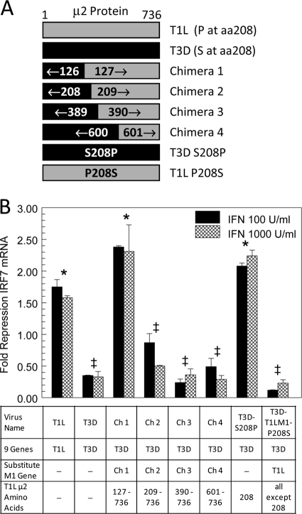
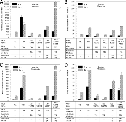
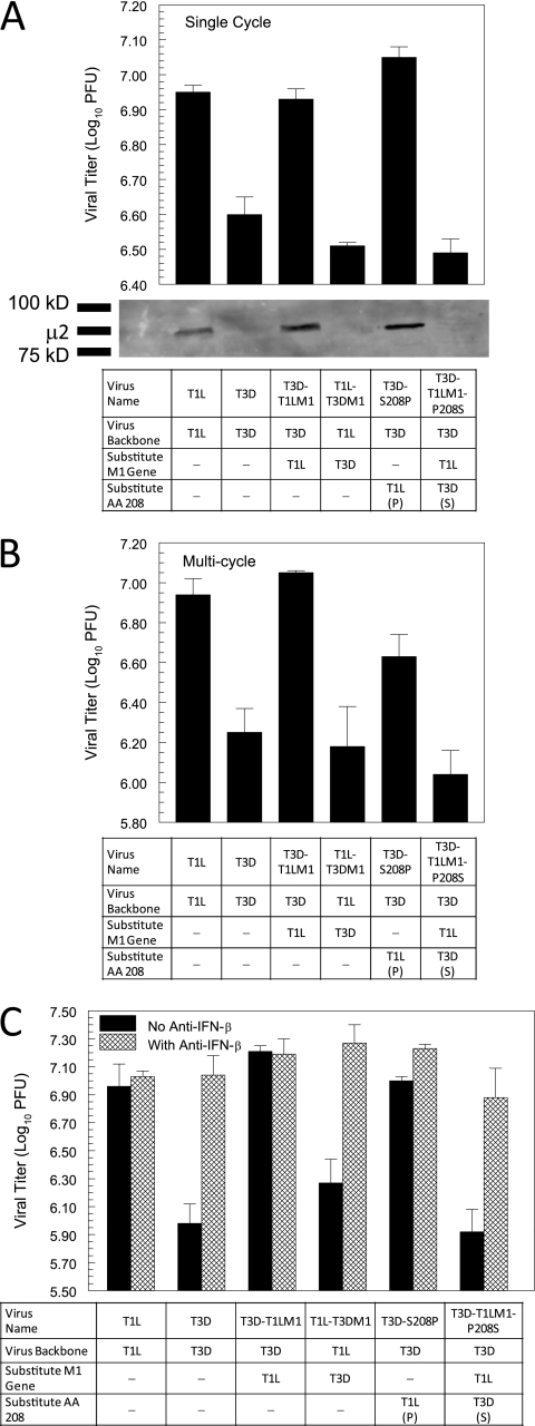
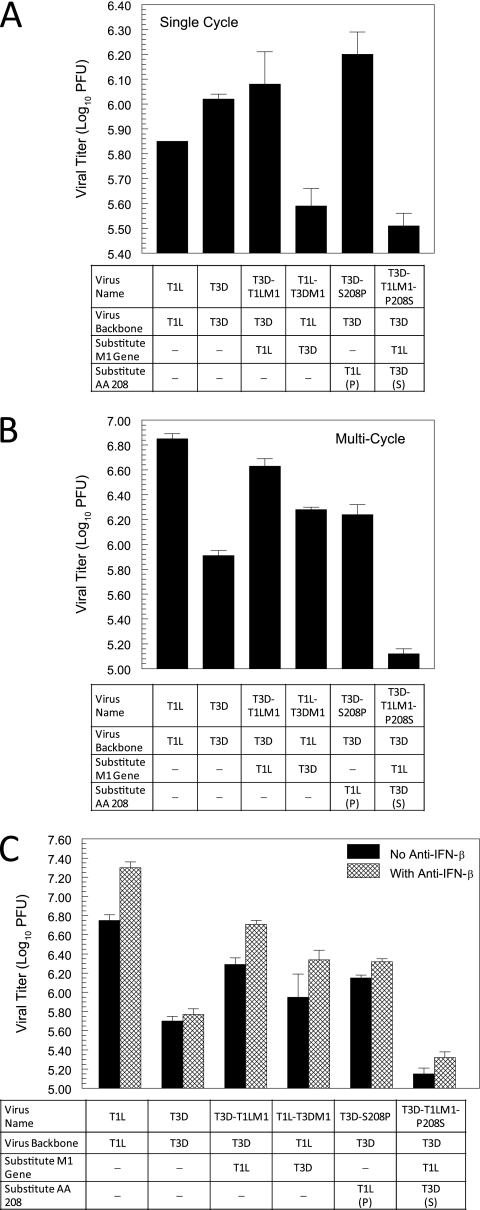
Myocarditis is indicated as the second leading cause of sudden death in young adults. Reovirus induces myocarditis in neonatal mice, providing a tractable model system for investigation of this important disease. Alpha/beta-interferon (IFN-α/β) treatment improves cardiac function and inhibits viral replication in patients with chronic myocarditis, and the host IFN-α/β response is a determinant of reovirus strain-specific differences in induction of myocarditis. Virus-induced IFN-β stimulates a signaling cascade that establishes an antiviral state and further induces IFN-α/β through an amplification loop. Reovirus strain-specific differences in induction of and sensitivity to IFN-α/β are associated with the viral M1, L2, and S2 genes. The reovirus M1 gene-encoded μ2 protein is a strain-specific repressor of IFN-β signaling, providing one possible mechanism for the variation in resistance to IFN and induction of myocarditis between different reovirus strains. We report here that μ2 amino acid 208 determines repression of IFN-β signaling and modulates reovirus induction of IFN-β in cardiac myocytes. Moreover, μ2 amino acid 208 determines reovirus replication, both in initially infected cardiac myocytes and after viral spread, by regulating the IFN-β response. Amino acid 208 of μ2 also influences the cytopathic effect in cardiac myocytes after spread. Finally, μ2 amino acid 208 modulates myocarditis in neonatal mice. Thus, repression of IFN-β signaling mediated by reovirus μ2 amino acid 208 is a determinant of the IFN-β response, viral replication and damage in cardiac myocytes, and myocarditis. These results demonstrate that a single amino acid difference between viruses can dictate virus strain-specific differences in suppression of the host IFN-β response and, consequently, damage to the heart.
Figures

Similar articles
-
Reovirus mu2 protein inhibits interferon signaling through a novel mechanism involving nuclear accumulation of interferon regulatory factor 9.J Virol. 2009 Mar;83(5):2178-87. doi: 10.1128/JVI.01787-08. Epub 2008 Dec 24. J Virol. 2009. PMID: 19109390 Free PMC article.
-
Reovirus induction of and sensitivity to beta interferon in cardiac myocyte cultures correlate with induction of myocarditis and are determined by viral core proteins.J Virol. 1998 Feb;72(2):1314-23. doi: 10.1128/JVI.72.2.1314-1323.1998. J Virol. 1998. PMID: 9445032 Free PMC article.
-
Basal and reovirus-induced beta interferon (IFN-beta) and IFN-beta-stimulated gene expression are cell type specific in the cardiac protective response.J Virol. 2005 Mar;79(5):2979-87. doi: 10.1128/JVI.79.5.2979-2987.2005. J Virol. 2005. PMID: 15709018 Free PMC article.
-
Pathogenesis of reovirus myocarditis.Curr Top Microbiol Immunol. 1998;233(Pt 2):51-66. doi: 10.1007/978-3-642-72095-6_3. Curr Top Microbiol Immunol. 1998. PMID: 9599931 Review. No abstract available.
-
The role of interferon regulatory factors in the cardiac response to viral infection.Viral Immunol. 2002;15(1):17-28. doi: 10.1089/088282402317340206. Viral Immunol. 2002. PMID: 11952139 Review.
Cited by
-
The μ2 and λ1 Proteins of Mammalian Reovirus Modulate Early Events Leading to Induction of the Interferon Signaling Network.Viruses. 2022 Nov 26;14(12):2638. doi: 10.3390/v14122638. Viruses. 2022. PMID: 36560642 Free PMC article.
-
Infectious factors in myocarditis: a comprehensive review of common and rare pathogens.Egypt Heart J. 2024 May 24;76(1):64. doi: 10.1186/s43044-024-00493-3. Egypt Heart J. 2024. PMID: 38789885 Free PMC article. Review.
-
De novo assembly of Sockeye salmon kidney transcriptomes reveal a limited early response to piscine reovirus with or without infectious hematopoietic necrosis virus superinfection.BMC Genomics. 2016 Nov 2;17(1):848. doi: 10.1186/s12864-016-3196-y. BMC Genomics. 2016. PMID: 27806699 Free PMC article.
-
Piscine orthoreovirus subtype 3 (PRV-3) causes heart inflammation in rainbow trout (Oncorhynchus mykiss).Vet Res. 2019 Feb 18;50(1):14. doi: 10.1186/s13567-019-0632-4. Vet Res. 2019. PMID: 30777130 Free PMC article.
-
Systems biology unravels interferon responses to respiratory virus infections.World J Biol Chem. 2014 Feb 26;5(1):12-25. doi: 10.4331/wjbc.v5.i1.12. World J Biol Chem. 2014. PMID: 24600511 Free PMC article. Review.
References
-
- Barnard P, McMillan NA. 1999. The human papillomavirus E7 oncoprotein abrogates signaling mediated by interferon-alpha. Virology 259:305–313 - PubMed
Publication types
MeSH terms
Substances
Grants and funding
LinkOut - more resources
Full Text Sources
