

doi: 10.1128/JVI.06223-11.
Epub 2011 Dec 7.
The Andes hantavirus NSs protein is expressed from the viral small mRNA by a leaky scanning mechanism
Affiliations
- PMID: 22156529
- PMCID: PMC3302399
- DOI: 10.1128/JVI.06223-11
Item in Clipboard
The Andes hantavirus NSs protein is expressed from the viral small mRNA by a leaky scanning mechanism
J Virol.
2012 Feb.
Abstract
The small mRNA (SmRNA) of all Bunyaviridae encodes the nucleocapsid (N) protein. In 4 out of 5 genera in the Bunyaviridae, the smRNA encodes an additional nonstructural protein denominated NSs. In this study, we show that Andes hantavirus (ANDV) SmRNA encodes an NSs protein. Data show that the NSs protein is expressed in the context of an ANDV infection. Additionally, our results suggest that translation initiation from the NSs initiation codon is mediated by ribosomal subunits that have bypassed the upstream N protein initiation codon through a leaky scanning mechanism.
Figures
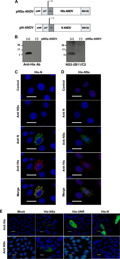
Detection of a transiently expressed recombinant His-ANDV NSs protein in Vero E6 cells. (A) Schematic representation of the plasmids used to express the recombinant tagged ADNV NSs and ANDV N proteins. The arrows indicate the initiation codon used to generate the recombinant proteins. BGH(A) corresponds to the bovine growth hormone polyadenylation sequences. (B) Plasmid pNSs-ANDV, expressing the recombinant His-ANDV NSs protein, was transfected in Vero E6 cells, and 48 h posttransfection total proteins were extracted. Shown is the expression of the recombinant His-ANDV NSs protein detected by Western blotting using a commercial anti-His monoclonal antibody (left) or the anti-NSs monoclonal antibody NS2-2B11/C2 (right). (C and D) Vero E6 cells transfected with plasmid pN-ANDV (C) or plasmid pNSs-ANDV (D) were fixed, and the expression of the His-tagged protein was detected by immunofluorescence using a commercial polyclonal anti-His antibody, anti-NSs monoclonal antibody NS2-5E7/D9, or a well-characterized anti-ANDV N monoclonal antibody (11, 30, 48). A goat anti-mouse fluorescein isothiocyanate (FITC)-conjugated antibody (green staining) and a goat anti-rabbit Alexa Fluor 555-conjugated antibody (red staining) were used as secondary antibodies. Cell nuclei were stained with 4′,6-diamidino-2-phenylindole (DAPI; blue staining). (E) Vero E6 cells transfected with pNSs-ANDV, pN-ANDV, and the His-tagged irrelevant tagged protein upstream of N-ras (His-UNR) were fixed, and the expression of the His-tagged proteins was detected by immunofluorescence using a commercial anti-His monoclonal antibody, followed by an FITC-labeled anti-mouse antibody (green staining). The recombinant His-tagged ANDV NSs protein was specifically detected by the anti-NSs monoclonal antibody NS2-5E7/D9, followed by an FITC-labeled anti-mouse antibody (green staining). Cell nuclei were stained with DAPI (blue staining). For all panels in the figure, size bars corresponds to 20 μm.
Detection of the ANDV NSs protein in infected Vero E6 cells. Vero E6 cells were infected with the ANDV CHI-7913 strain as previously described (10, 11, 30). At 6, 12, 24, and 48 h postinfection (p.i.), cells were fixed and the presence of the ANDV NSs protein was evaluated by IF using antibody NS2-5E7/D9, followed by an FITC-labeled anti-mouse secondary antibody (green staining). In parallel, the ANDV N protein was detected using a well-characterized polyclonal antibody (10) followed by an Alexa Fluor 555-labeled anti-rabbit secondary antibody (red staining). The cellular nucleus was stained with DAPI (blue staining). Size bars correspond to 20 μm. (B) Total RNA was extracted from ANDV-infected cells 2 (lane 2), 6 (lane 3), 12 (lane 4), and 24 h (lane 5) postinfection (h.p.i) and used as the template in an RT-PCR designed to specifically amplify the viral S-RNAs (genomic S-RNA, smRNA, and the positive-sense antigenome scRNA) (11, 30). This assay also included a positive RT-PCR control that consisted of RNA extracted from the supernatant initially used to infect Vero E6 cells (viral stock, lane 7) and a negative RT-PCR control which corresponds to water (lane 8). MW indicates the molecular size marker (1 kb; lane 1; Fermentas).
NSs ORF can be translated in the context of virus-like smRNAs. (A) Schematic representation of the capped monocistronic RNA constructs that mimic the ANDV S mRNA. To faithfully mimic the viral mRNA, these RNAs contain 21 additional nucleotides (5′-GGGAGACCCAAGCTGGCTAGC-3′; gray region) between the 5′ cap structure (oval at the 5′ end of the RNA) and the first nucleotide of the ANDV smRNA sequence (black line) (50). Additionally, the viral NSs ORF has been replaced by the firefly luciferase (FLuc) reporter gene. The arrow indicates the initiation codon used. In the N RNA, the N initiation codon is in frame with FLuc. In the NSs RNA, the putative NSs initiation codon is in frame with FLuc. In the CUC RNAs, the AUGN or AUGNSs codons have been mutated to CUC. (B) The N RNA (black bar) and NSs RNA (gray bar) were translated in a commercial in vitro translation system as indicated in Materials and Methods. FLuc activity (in relative light units [RLU]) was measured as indicated in Materials and Methods. Unshaded bars correspond to FLuc activity obtained with CUC mutations. Values are the means ± standard deviations (SD) from three independent experiments, each conducted in triplicate. (C) DNA plasmids coding for the RNAs depicted in panel A plus an RLuc-expressing plasmid (transfection efficiency control) were transfected into HeLa cells. Total RNA and protein were extracted and analyzed. RNA was used to assess the total amount of the virus-like RNAs in cells by an RT-qPCR assay (right panel). The quantity of N RNA was arbitrarily set to 1, and the total amount of NSs RNA recovered from cells was expressed relative to this value (right panel). FLuc and RLuc activities were determined, and FLuc activity was normalized to RLuc activity (left panel). Normalized N RNA FLuc activity was arbitrarily set to 1 (left panel). Values shown in both panels are the means ± SD from three independent experiments each conducted in triplicate.

Translation initiation from the N and NSs initiation codons is sensitive to edeine. (A) Schematic representation of a bicistronic mRNA similar to those used in this study. RNAs correspond to the dual-luciferase (dl) reporter construct containing an upstream Renilla luciferase gene (RLuc) and a downstream firefly luciferase gene (FLuc). IRES activity was monitored using the FLuc activity as the readout, while the RLuc reporter gene served as a control for cap-dependent translation initiation. Two dl RNAs were used in this study, one harboring the poliovirus (PV) IRES (37) and the other harboring the hepatitis C virus (HCV) IRES (1). dl PV (B) or dl HCV (C) bicistronic RNA was translated in RRL in the presence of the translation initiation inhibitor edeine, which interferes with AUG start codon recognition by scanning eIF2-GTP/Met-tRNAiMet complexes (7, 14, 25, 26). The relative RLuc and FLuc activity for each RNA in the absence of edeine was arbitrarily set to 1 (± SD). Values are the means ± SD from three independent experiments each conducted in triplicate. (D) Monocistronic ANDV S-like mRNAs depicted in Fig. 1A were translated in RRL in the presence of edeine. The relative FLuc activity for each RNA in the absence of edeine was arbitrarily set to 1 (± SD). Values are the means ± SD from at least three independent experiments, each conducted in triplicate.

Sequence context of the 5′-proximal N initiation codon modulates the recognition of the putative NSs initiation codon. RNAs corresponding to different mutants of the NSs RNA (Table 2) were translated in RRL (A), or plasmid DNA coding for them was transfected into HeLa cells together with an RLuc-expressing plasmid (as a control for the transfection efficiency) (B). W stands for the wild-type context as found in the ADNV smRNA, K stands for the optimal context (purine in position −3 and a G in position +4), and in X the AUG initiation codon has been mutated to CUC. FLuc values were normalized to RLuc activity. The relative FLuc activity for the wild-type (W/W) virus-like RNA was arbitrarily set to 1 (± SD). Values are the means ± SD from three independent experiments each conducted in triplicate.
References
Publication types
MeSH terms
Substances
Grants and funding
LinkOut - more resources
Full Text Sources
Other Literature Sources
Medical
