. 2012 Feb;86(4):1999-2010.
doi: 10.1128/JVI.05871-11. Epub 2011 Dec 7.

Evolution of poliovirus defective interfering particles expressing Gaussia luciferase

Affiliations

Evolution of poliovirus defective interfering particles expressing Gaussia luciferase

Yutong Song et al. J Virol. 2012 Feb.

Abstract

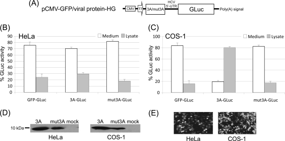
Polioviruses (PVs) carrying a reporter gene are useful tools for studies of virus replication, particularly if the viral chimeras contain the polyprotein that provides all of the proteins necessary for a complete replication cycle. Replication in HeLa cells of a previously constructed poliovirus expressing the gene for Renilla luciferase (RLuc) fused to the N terminus of the polyprotein H(2)N-RLuc-P1-P2-P3-COOH (P1, structural domain; P2 and P3, nonstructural domains) led to the deletion of RLuc after only one passage. Here we describe a novel poliovirus chimera that expresses Gaussia luciferase (GLuc) inserted into the polyprotein between P1 and P2 (N(2)H-P1-GLuc-P2-P3-COOH). This chimera, termed PV-GLuc, replicated to 10% of wild-type yield. The reporter signal was fully retained for three passages and then gradually lost. After six passages the signal was barely detectable. On further passages, however, the GLuc signal reappeared, and after eight passages it had reached the same levels observed with the original PV-GLuc at the first passage. We demonstrated that this surprising observation was due to coevolution of defective interfering (DI) particles that had lost part or all of the capsid coding sequence (ΔP1-GLuc-P2-P3) and wild-type-like viruses that had lost the GLuc sequence (P1-P2-P3). When used at low passage, PV-GLuc is an excellent tool for studying aspects of genome replication and morphogenesis. The GLuc protein was secreted from mammalian cells but, in agreement with published data, was not secreted from PV-GLuc-infected cells due to poliovirus-induced inhibition of cellular protein secretion. Published evidence indicates that individual expression of enterovirus polypeptide 3A, 2B, or 2BC in COS-1 cells strongly inhibits host protein secretion. In HeLa cells, however, expression of none of the poliovirus polypeptides, either singly or in pairs, inhibited GLuc secretion. Thus, inhibition of GLuc secretion in PV-infected HeLa cells is likely a result of the interaction between several viral and cellular proteins that are different from those in COS-1 cells.


Figures

Fig 1
Fig 1
Characterization of novel monocistronic reporter construct pPV-GLuc. (A) Schematic representation of the GLuc-containing reporter construct. The polioviral reporter genome contains the coding sequence for GLuc inserted between capsid precursor P1 and the nonstructural P2 domain, followed by the FMDV 2A peptide flanked by two different in-frame clone linkers. Solid and dotted arrows, cleavage sites by PV 2A and FMDV 2A, respectively; shaded box followed by the 2A cleavage site at the N terminus of the polyprotein, a linker of 27 nt encoding the sequence ENLTTY. (B) One-step growth curves of wt PV1(M) and of the reporter virus PV-GLuc. HeLa cell monolayers were infected with viral supernatants of wt PV and of PV-GLuc (passage 1), and the virus yield was determined at the indicated time points by plaque assay (see Materials and Methods). (C) Number of RNA genome copies in PV1(M)- and PV GLuc-infected HeLa cells. The number of RNA genomes was measured at various times postinfection by quantitative RT-PCR (see Materials and Methods). (D) GLuc activity both in the growth medium and in the lysate was determined at the indicated time points postinfection.
Fig 2
Fig 2
Growth properties of PV-GLuc. (A) Summary of total GLuc activities at different viral passages. All passages were carried out in the absence or presence of GnHCl (2 mM). GLuc measurements were made at between 18 and 20 h postinfection at the time when cytopathic effects were observed. (B) Plaque phenotypes of wt PV and of PV-GLuc at passage 1 (P1) and passage 8 (P8) (see Materials and Methods).
Fig 3
Fig 3
Genetic stability of PV-GLuc. (A) Genetic stability of PV-GLuc during passaging on HeLa cells. Viral RNA was reverse transcribed, followed by PCR, and the length of the cDNAs was analyzed on an agarose gel. Lanes M, DNA 1-kb marker (NEB); lane 1, PCR fragment derived from the entire P1 and GLuc coding sequences of PV-GLuc; lanes 2 and 3, PCR fragments derived from PV-GLuc RNA isolated after passages 3 and 4, respectively; lane 4, control PCR fragment derived from wt poliovirus RNA. (B) Identification of DI particles derived from PV-GLuc. Viral RNAs obtained at different passages were reverse transcribed, and following PCR, the DNA fragments obtained were analyzed on an agarose gel. Lanes M, DNA 1-kb marker (NEB); lane 1, negative control (no RNA); lane 2, PCR fragment derived from the entire P1 and GLuc coding sequences of pPV-GLuc; lane 3, PCR fragment derived from wt poliovirus RNA; lanes 4 to 6, PCR fragments derived from PV-GLuc RNA isolated at passages 6, 9, and 21, respectively. (C) Quantitation of helper virus and of DI genomes. The percentage of helper virus and of DI genomes in cell lysates obtained at passages 8, 9, and 21 was determined by quantitative RT-PCR (see Materials and Methods).
Fig 4
Fig 4
Genetic analysis of DI particles derived from PV-GLuc and interference with wt poliovirus growth by DI genomes. (A) Schematic diagram of PV-GLuc. The region from nt 626 to nt 4148 is enlarged in panel B. Arrows, location of primers used for RT-PCR. (B) Viral or DI RNAs were reverse transcribed, and following PCR amplification of the region from nt 626 to nt 4148, the DNA fragments were sequenced. Solid black lines, sequences retained from the parental PV-GLuc in the helper virus or the DI genomes; waved gray lines, GLuc coding sequences; dotted lines, deleted regions. Numbers above the lines indicate the position in the parental pPV-GLuc plasmid of the last upstream nucleotide before and the first downstream nucleotide after each deletion. On the right, the passage number from which the sample was derived is indicated. The total number of samples sequenced is indicated in parentheses after the passage number. DI genome 12, labeled with an asterisk, is derived from the CsCl gradient separation. (C) Virus titers at different passages (passages 1, 6, 9, and 21) of PV-GLuc on HeLa cells were determined by plaque assay (Materials and Methods).
Fig 5
Fig 5
Effect of 3A expression on secretion of GLuc in HeLa and COS-1 cells. (A) Schematic representation of the dicistronic expression constructs used in this study. The first cistron contains the coding sequence either of 3A (wt or mutant 3A [mut3A]) or of a control protein (eGFP). The second cistron contains the GLuc coding sequence preceded by the HCV IRES. UTR, untranslated region. (B) Effect of 3A (wt or mutant 3A) or control protein expression on the secretion of GLuc in HeLa cells. HeLa cells were transfected with the dicistronic reporter DNA plasmids. GLuc activities, both secreted and intracellular, were measured at about 24 h posttransfection. The data are plotted as a percentage of the total GLuc activity. Error bars indicate the standard deviations of measurements from triplicate experiments. (C) Effect of 3A (wt or mutant 3A) or control protein expression on the secretion of GLuc in COS-1 cells. The experiment was the same as described for panel B, except that COS-1 cells were used. (D) Expression of wt 3A or mutant 3A proteins in HeLa and COS-1 cells. The expression of 3A proteins was monitored by Western blot analysis of samples collected at 24 h posttransfection. (E) Expression of eGFP in HeLa and COS-1 cells at 24 h posttransfection visualized by fluorescence microscopy. The efficiency of transfection was determined by flow cytometry: 51% of HeLa cells and 61% of COS-1 cells were transfected.
Fig 6
Fig 6
Effect of expression of viral proteins 2B, 2C, 2BC, and 3AB individually on the secretion of GLuc in HeLa and COS-1 cells. (A) Schematic representation of the dicistronic constructs used in the study. The first cistron contained either the viral or the control (eGFP) protein, while the second cistron contained the GLuc coding sequences. (B) Effect of viral or control protein expression on the secretion of GLuc in HeLa cells. HeLa cells were transfected with the dicistronic reporter DNA plasmids. GLuc activities, both secreted and intracellular, were measured at about 24 h posttransfection. The data are plotted as a percentage of the total GLuc activity. Error bars indicate the standard deviations of measurements from duplicate experiments. (C) Effect of viral or control protein expression on the secretion of GLuc in COS-1 cells. The experiment was the same as described for panel B, except that COS-1 cells were used. (D) Expression of viral proteins in HeLa and COS-1 cells was monitored by Western blot analysis of samples collected at 24 h posttransfection.
Fig 7
Fig 7
Effect of coexpression of 3A with another viral protein (2B, 2C, or 2BC) on the secretion of GLuc in HeLa and COS-1 cells. (A) Schematic representation of the tricistronic constructs used in the study. In the first cistron, translation of PV 3A is initiated by a cap-dependent mechanism. The second cistron contained either a viral protein (2B, 2C, or 2BC) or the control (eGFP) protein, while the third cistron contained the GLuc coding sequences. (B) Effect of viral or control protein coexpression on the secretion of GLuc in HeLa cells. HeLa cells were transfected with the tricistronic reporter DNA plasmids. GLuc activities, both secreted and intracellular, were measured at about 24 h posttransfection. The data are plotted as a percentage of the total GLuc activity. Error bars indicate the standard deviations of measurements from duplicate experiments. (C) Effect of viral or control protein coexpression on the secretion of GLuc in COS-1 cells. The experiment was the same as described for panel B, except that COS-1 cells were used. (D) Expression of viral proteins in HeLa and COS-1 cells was monitored by Western blot analysis of samples collected at 24 h posttransfection.
Fig 8
Fig 8
Effect of brefeldin A on GLuc secretion. (A) Schematic representation of the GLuc-expressing reporter construct pCMV-GLuc. (B) Inhibition of GLuc transport in the presence of the indicated concentrations of BFA. HeLa cells were transfected with a GLuc-expressing vector. At 6 h posttransfection, cells were treated with different amounts of BFA. Samples taken from growth medium and lysates were assayed for GLuc activity at 16 h after addition of BFA.

Similar articles

Cited by

References

    1. Agol VI, Gmyl AP. 2010. Viral security proteins: counteracting host defences. Nat. Rev. Microbiol. 8:867–878 - PMC - PubMed
    1. Aldabe R, Barco A, Carrasco L. 1996. Membrane permeabilization by poliovirus proteins 2B and 2BC. J. Biol. Chem. 271:23134–23137 - PubMed
    1. Alexander L, Lu HH, Gromeier M, Wimmer E. 1994. Dicistronic polioviruses as expression vectors for foreign genes. AIDS Res. Hum. Retroviruses 10(Suppl 2):S57–S60 - PubMed
    1. Andino R, Rieckhof GE, Achacoso PL, Baltimore D. 1993. Poliovirus RNA synthesis utilizes an RNP complex formed around the 5′-end of viral RNA. EMBO J. 12:3587–3598 - PMC - PubMed
    1. Badr CE, Hewett JW, Breakefield XO, Tannous BA. 2007. A highly sensitive assay for monitoring the secretory pathway and ER stress. PLoS One 2:e571. - PMC - PubMed

Publication types