. 2012 Mar 15;21(6):1384-90.
doi: 10.1093/hmg/ddr576. Epub 2011 Dec 8.

A critical role for the PAR-1/MARK-tau axis in mediating the toxic effects of Aβ on synapses and dendritic spines

Affiliations

A critical role for the PAR-1/MARK-tau axis in mediating the toxic effects of Aβ on synapses and dendritic spines

Wendou Yu et al. Hum Mol Genet. .

Abstract

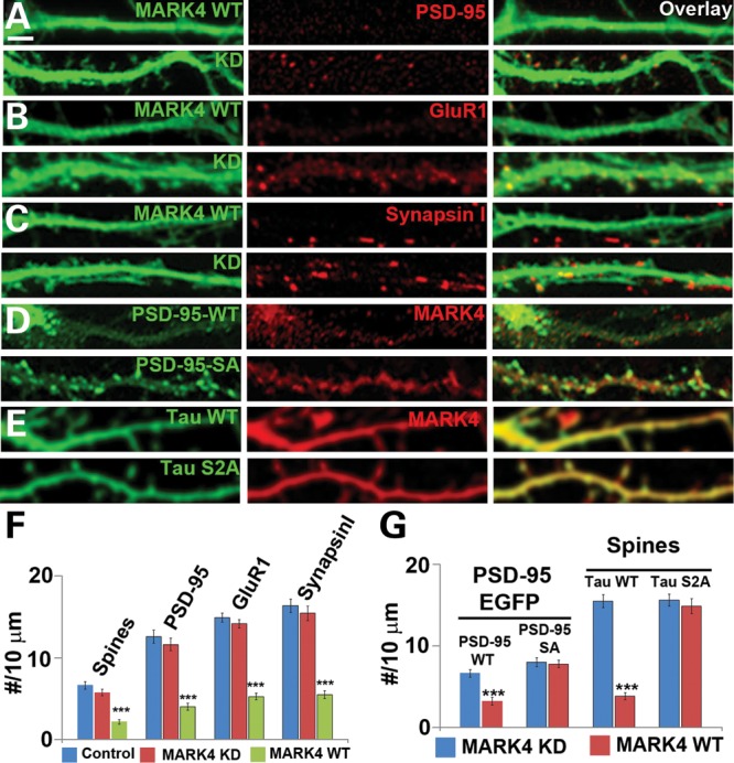
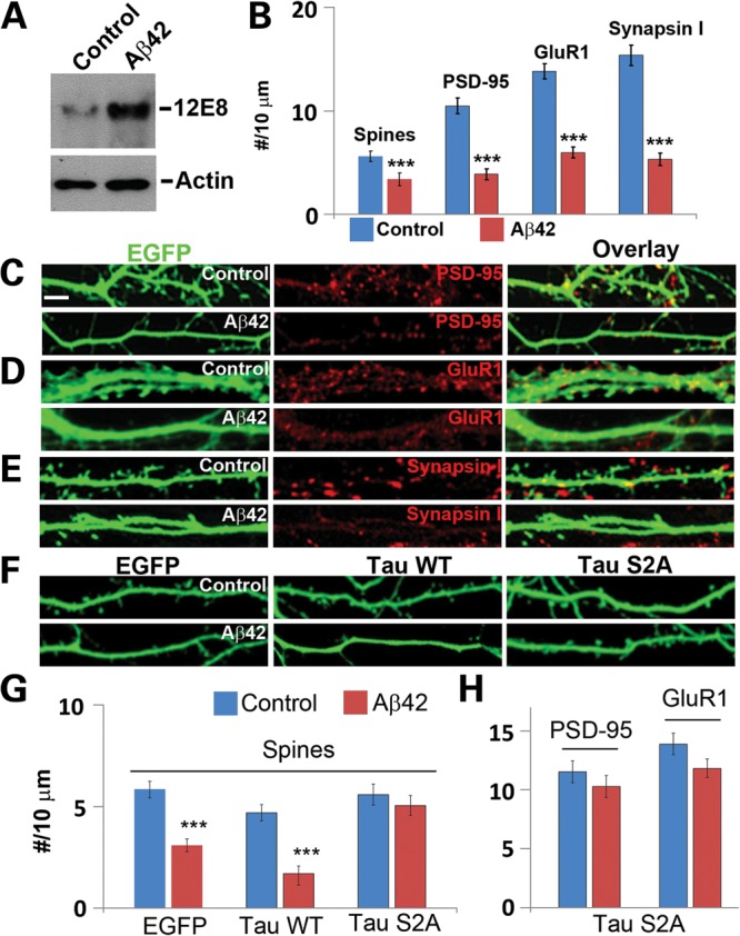
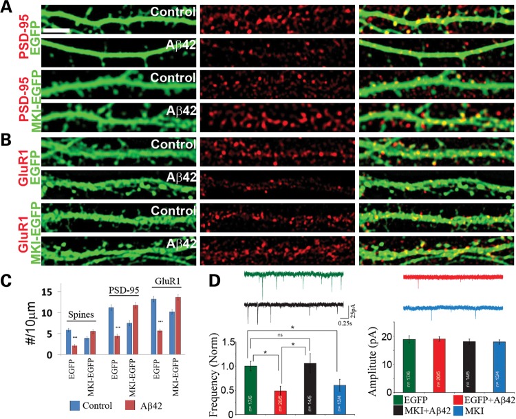
Alzheimer's disease (AD) is the most common neurodegenerative disease and the leading cause of dementia in the elderly. Accumulating evidence supports soluble amyloid-β (Aβ) oligomers as the leading candidate for the causative agent in AD and synapses as the primary site of Aβ oligomer action. However, the molecular and cellular mechanisms by which Aβ oligomers cause synaptic dysfunction and cognitive impairments remain poorly understood. Using primary cultures of rat hippocampal neurons as a model system, we show that the partitioning defective-1 (PAR-1)/microtubule affinity-regulating kinase (MARK) family kinases act as critical mediators of Aβ toxicity on synapses and dendritic spines. Overexpression of MARK4 led to tau hyperphosphorylation, reduced expression of synaptic markers, and loss of dendritic spines and synapses, phenotypes also observed after Aβ treatment. Importantly, expression of a non-phosphorylatable form of tau with the PAR-1/MARK site mutated blocked the synaptic toxicity induced by MARK4 overexpression or Aβ treatment. To probe the involvement of endogenous MARK kinases in mediating the synaptic toxicity of Aβ, we employed a peptide inhibitor capable of effectively and specifically inhibiting the activities of all PAR-1/MARK family members. This inhibitor abrogated the toxic effects of Aβ oligomers on dendritic spines and synapses as assayed at the morphological and electrophysiological levels. Our results reveal a critical role for PAR-1/MARK kinases in AD pathogenesis and suggest PAR-1/MARK inhibitors as potential therapeutics for AD and possibly other tauopathies where aberrant tau hyperphosphorylation is involved.


Figures

Figure 1.
Figure 1.
Overexpression of MARK4 causes synaptic and spine abnormality in rat hippocampal neurons. (AC) Effects of MARK4-WT or MARK4-KD overexpression on postsynaptic PSD-95 clusters (A), AMPA receptor GluR1 clusters (B), presynaptic Synapsin I clusters (C) and spine numbers (A–C). Data quantification is shown in (F). (D) Differential response of PSD-95-S561A and PSD-95-WT synaptic localization to MARK4-WT overexpression. (E) Effect of h-tau-WT or h-tau-S2A co-expression on MARK4-WT overexpression-induced spines loss. (F) Quantification of spine number and PSD-95 (A), GluR1 (B) and Synapsin I (C) clusters in neurons transfected with MARK4-WT or MARK4-KD (***P< 0.001 in Student's t-test). (G) (Left) Quantification of PSD-95 clusters in neurons co-transfected with MARK4-WT or MARK4-KD and EGFP-tagged PSD-95-WT or PSD-95-S561A (***P< 0.001 in Student's t-test). (Right) Quantification of spine number in neurons co-transfected with MARK4-WT or MARK4-KD and h-tau-WT or h-tau-S2A (***P< 0.001 in Student's t-test). Scale bar, 5 μm.
Figure 2.
Figure 2.
Synaptic and spine toxicity induced by Aβ oligomers and rescue of Aβ-induced spine loss by h-tau-S2A. (A) Aβ treatment increased endogenous tau phosphorylation at the 12E8 sites. Hippocampal neurons at 13 days in vitro were treated with Aβ oligomers for 12h and used for western blot analysis. Actin serves as loading control. (BE) Effects of Aβ treatment on PSD-95 clusters (C), GluR1 clusters (D), Synapsin I clusters (E) and spine number (B) in EGFP-transfected neurons. Data quantification is shown in (B) (***P< 0.001 in Student's t-test). (FH) Resistance of h-tau-S2A-transfected neurons to Aβ-induced loss of spines (F and G) and PSD-95 and GluR1 clusters (H). Scale bar, 10 μm.
Figure 3.
Figure 3.
Inhibition of PAR-1/MARK-mediated tau phosphorylation and MARK4 overexpression-induced synaptic and spine toxicity by MKI. (A) MKI-EGFPinhibited phosphorylation of endogenous tau as assessed by western blot analysis using phosphor-specific antibodies 12E8 and PHF-1 (A). (B) MKI-EGFP inhibited phosphorylation of HA-tagged exogenous h-tau-WT at the 12E8 sites. Robust 12E8 staining (red) is seen in EGFP-transfected control neurons. Total tau is stained with anti-HA (blue). (C) No effect of MKI-EGFP on AMPK kinase activity as detected with the phospho-acetyl-CoA carboxylase (Ser79) (pACC) antibody. EGFP-transfected neurons serve as control. (D and E) MKI-EGFP blocked MARK4-overexpression-induced loss of spines (D and E) and PSD-95 and GluR1 clusters (E). Data quantification is shown in (E) (***P< 0.001 in Student's t-test). Scale bar, 10 μm.
Figure 4.
Figure 4.
Rescue of Aβ-induced synaptic and dendritic spine defects by MKI at the morphological and electrophysiological levels. (AC) Effects of MKI-EGFP on Aβ-induced loss of PSD-95 (A) and GluR1 (B) clusters and spine numbers. Neurons transfected with MKI-EGFP or EGFP were treated with Aβ oligomer or mock-treated with solvent as control. The cytoplasmic GFP signals allowed detection of dendritic spines, and PSD-95 or GluR1 clusters were detected by immunostaining. Quantification of data is shown in (C) (***P< 0.001 in Student's t-test). (D) Sample traces of synaptic activity (mEPSCs) recorded in neurons from four different conditions (control-EGFP, Aβ, MKI-EGFP + Aβ and MKI-EGFP alone) held at −70 mV. Summary graphs from four to six culture sets plotting normalized mEPSC frequency and mEPSC amplitude are shown beneath the traces. Scale bar, 5 μm.

Similar articles

Cited by

References

    1. Selkoe D.J. Alzheimer's disease is a synaptic failure. Science. 2002;298:789–791. - PubMed
    1. Penzes P., Cahill M.E., Jones K.A., VanLeeuwen J.E., Woolfrey K.M. Dendritic spine pathology in neuropsychiatric disorders. Nat. Neurosci. 2011;14:285–293. - PMC - PubMed
    1. Spires T.L., Meyer-Luehmann M., Stern E.A., McLean P.J., Skoch J., Nguyen P.T., Bacskai B.J., Hyman B.T. Dendritic spine abnormalities in amyloid precursor protein transgenic mice demonstrated by gene transfer and intravital multiphoton microscopy. J. Neurosci. 2005;25:7278–7287. - PMC - PubMed
    1. Reddy P.H., Beal M.F. Amyloid beta, mitochondrial dysfunction and synaptic damage: implications for cognitive decline in aging and Alzheimer's disease. Trend. Mol. Med. 2008;14:45–53. - PMC - PubMed
    1. Selkoe D.J. Soluble oligomers of the amyloid beta-protein impair synaptic plasticity and behavior. Behav. Brain Res. 2008;192:106–113. - PMC - PubMed

Publication types

MeSH terms