. 2012 Jan 15;188(2):724-34.
doi: 10.4049/jimmunol.1102630. Epub 2011 Dec 12.

Loss of B7-H1 expression by recipient parenchymal cells leads to expansion of infiltrating donor CD8+ T cells and persistence of graft-versus-host disease

Affiliations

Loss of B7-H1 expression by recipient parenchymal cells leads to expansion of infiltrating donor CD8+ T cells and persistence of graft-versus-host disease

Xiaofan Li et al. J Immunol. .

Abstract

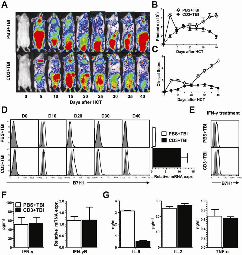
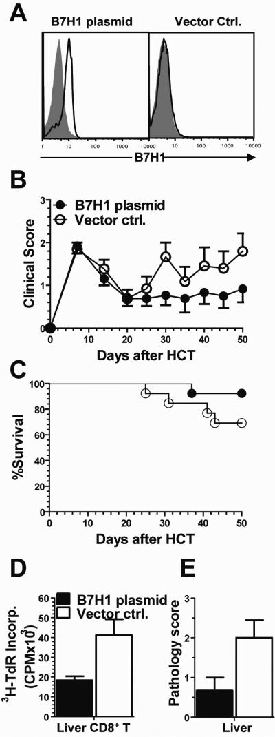
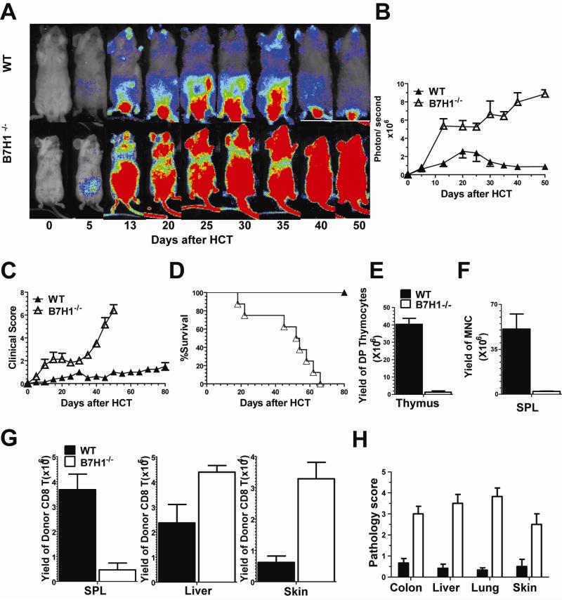
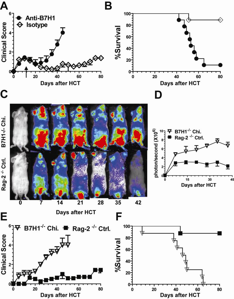
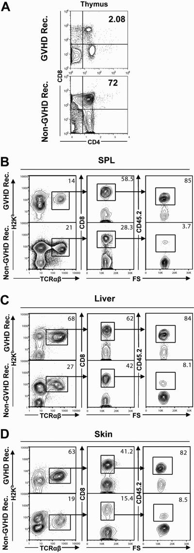
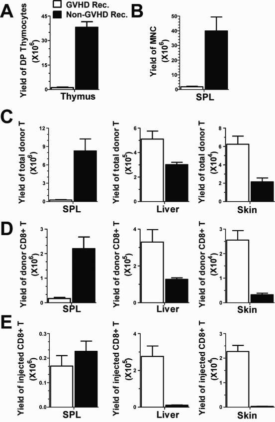
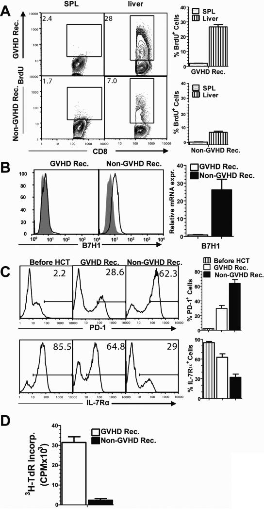
Previous experimental studies have shown that acute graft-versus-host disease (GVHD) is associated with two waves of donor CD8(+) T cell expansion. In the current studies, we used in vivo bioluminescent imaging, in vivo BrdU labeling, and three different experimental GVHD systems to show that B7-H1 expression by recipient parenchymal cells controls the second wave of alloreactive donor CD8(+) T cell expansion and the associated second phase of GVHD. Loss of B7-H1 expression by parenchymal cells during the course of GVHD was associated with persistent proliferation of donor CD8(+) T cells in GVHD target tissues and continued tissue injury, whereas persistent expression of B7-H1 expression by parenchymal cells led to reduced proliferation of donor CD8(+) T cells in GVHD target tissues and resolution of GVHD. These studies demonstrate that parenchymal cell expression of B7-H1 is required for tolerizing infiltrating T cells and preventing the persistence of GVHD. Our results suggest that therapies designed to preserve or restore expression of B7-H1 expression by parenchymal tissues in the recipient could prevent or ameliorate GVHD in humans.


Figures

Fig. 1
Fig. 1. Loss of B7-H1 expression by parenchymal tissues in TBI-conditioned recipients was associated with chronic inflammation
WT BALB/c recipients were pretreated with PBS or anti-CD3 mAb (5 μg/g) seven days before transplantation. On day 0, 8 hours after 800 cGy TBI, CD8+ T cells (20×106) from luc+ C57BL/6 donors and BM cells (50×106) from WT C57BL/6 donors were injected i.v. into recipients. Recipients were monitored for clinical GVHD 1-2 times a week. In vivo donor CD8+ T cell expansion was visualized with in vivo BLI every 5 days for up to 40 days after transplantation. (A) A representative series of in vivo BLI pictures depicting donor CD8+ T expansion is shown for 1 of 8 recipients in each group. (B) Kinetic curves of whole body emission of photons per second (mean ± SE, N=8). (C) Clinical GVHD scores (mean ± SE, N=8). (D) On days 0, 10, 20, 30, and 40 after transplantation, hepatocytes from the recipients with or without anti-CD3 pretreatment were isolated and stained with anti-B7-H1or isotype control. One representative FACS pattern is shown of three replicate experiments. Hepatocytes from D40 were also used for real-time PCR of B7-H1 mRNA. Relative expression levels of B7-H1 mRNA is shown (mean ± SE, N=4). (E) On day 30 after transplantation, recipients with or without anti-CD3 pretreatment were given 150 μg recombinant murine IFN-γ by i.v. injection. 48 hours later, hepatocytes from the recipients were stained with anti-B7-H1 or isotype control. One representative FACS pattern is shown of three replicate experiments. (F-G) On day 30 after transplantation, the infiltrating mononuclear cells (0.2×10⁶/well) from liver tissues were stimulated with PMA (10ng/ml) and ionomycin (200ng/ml) for 72 hours, and then the culture supernatants were measured with ELISA. (F) left panel: IFN-γ concentration (mean ± SE, N=4); right panel: relative expression levels of IFN-γR mRNA. (G) IL-6, IL-2, TNF-α concentration (mean ± SE, N=4).
Fig. 2
Fig. 2. Induction of hepatocyte expression of B7-H1 via hydrodynamic injection of B7-H1 cDNA inhibited tissue infiltrating donor CD8+ T cell expansion
WT BALB/c recipients were conditioned with 800cGy TBI and then given CD8+ T cells (5 × 10⁶) and BM cells (50 × 106) from WT C57BL/6 donors. 20 days after transplantation, the recipients were given hydrodynamic injection of B7-H1 cDNA or vector control. Thereafter, the recipients were monitored for clinical GVHD. (A) 24 hours after hydrodynamic injection, hepatocytes from recipients treated with B7-H1 plasmid or vector control were stained for anti-B7-H1 or IgG isotype control. A representative FACS pattern is shown from one of three replicate experiments. (B) Clinical GVHD scores (mean ± SE, N=13). (C) Survival curves (N=13). (D) 50 days after transplantation, sorted donor CD8+ T cells (0.2 ×10⁶/well) from liver tissues of recipients treated with B7-H1 plasmid or vector control were stimulated with immobilized anti-CD3 for 72 hours, and 3H-TdR incorporation was measured. Mean ± SE of CPM of 3H-TdR incorporation from 4 replicate experiments is shown. (E) 50 days after transplantation, liver tissue was harvested for histopathologic evaluation. The mean ± SE of pathology scores of 6 recipients per group are shown.
Fig. 3
Fig. 3. Expression of B7-H1 by recipient tissues prevented the second wave of donor CD8+ T cell expansion and the second phase of GVHD
WT BALB/c or B7-H1−/− recipients were conditioned with anti-CD3-based regimen and then given CD8+ T cells (20×106) from luc+ C57BL/6 donors and BM cells (50×106) from WT C57BL/6 donors. Recipients were monitored for clinical GVHD 1-2 times a week. Donor CD8+ T cell expansion was visualized with in vivo BLI for more than 50 days after transplantation. Additional recipients were used to compare donor T cell yield in lymphoid tissues and GVHD target tissues and to evaluate histopathology. (A) A representative series of in vivo BLI pictures depicting donor CD8+ T expansion is shown for 1 of 8 recipients in each group. (B) Kinetic curves of whole body emission of photons per second (mean ± SE, N=8). (C, D) Clinical GVHD score (mean ± SE) and survival curves, N=8. (E-G) Additional recipients were used for enumerating thymocytes, MNC in spleen, and donor T cells in spleen, liver, and skin via staining of H-2Kb, TCRβ, CD4 and CD8, 35 days after transplantation. (E) Yields of CD4+CD8+ thymocytes (mean ± SE, N=4). (F) Yields of MNCs in spleen (mean ± SE, N=4). (G) Yields of donor CD8+ T cells in spleen, liver and skin (mean ± SE, N=4). (H) Histopathology scores (mean ± SE, N=6).
Fig. 4
Fig. 4. Expression of B7-H1 by parenchymal tissues prevented the second wave of donor CD8+ T cell expansion and the second phase of GVHD in Rag-2−/− recipients
Rag2−/− BALB/c mice were given lethal TBI (800 cGy) and then given BM cells (5×10⁶) from B7-H1−/− Rag2−/− BALB/c mice to create B7-H1+/+ chimeras that had B7-H1 expression only in parenchymal cells. Conversely, B7-H1−/− chimeras with expression of B7-H1 in hematopoietic cells but not in parenchymal cells were established by transferring BM cells from Rag-2−/− mice into B7-H1−/−Rag2−/− mice. The control Rag-2−/− mice were given TBI and BM cells from Rag-2−/− donors. 60 days later, the B7-H1+/+ chimeras and the B7-H1−/− chimeras were used as transplantation recipients without conditioning. (A-B) B7-H1−/− chimeras were given CD8+ T cells (20 × 106) and BM cells (50 × 106) from C57BL/6 donors.14 days after transplantation, the recipients were given i.p. injections of anti-B7-H1 mAb or control rat IgG (5 μg/g) every other day for 30 days. Recipients were monitored for clinical GVHD and survival. (A) Kinetic curves of clinical score (N=9). (B) Survival curves (N=9). (C-F) B7-H1−/− chimeras and control Rag-2−/− mice were given luc+CD8+ T cells (20 × 106) from luciferase-transgenic C57BL/6 donors and BM cells (50 × 106) from WT C57BL/6 donors. Thereafter, recipients were monitored for clinical GVHD once or twice weekly. Donor CD8+ T cell expansion was visualized with in vivo BLI for more than 40 days after transplantation. (C) A representative series of in vivo BLI pictures depicting donor CD8+ T expansion is shown for 1 of 8 recipients in each group. (D) Kinetic curves of whole body emission of photons per second (N=8). (E) Clinical GVHD scores (N=8). (F) Survival curves (N=8).
Fig. 5
Fig. 5. Comparison of percentage of CD4+CD8+ thymocytes and percentage of injected donor CD8+ T cells in spleen and GVHD target tissues of recipients with and without GVHD
WT BALB/c recipients were conditioned with 800 cGy TBI or with anti-CD3, vorinostat, and busulfan, and then given CD8+ T cells (20×106) from WT (CD45.2) C57BL/6 donors and TCD-BM cells (50×106) from congenic (CD45.1) C57BL/6 donors. 35 days after transplantation, when TBI-conditioned recipients showed severe clinical GVHD, the percentage of CD4+CD8+ thymocytes and percentage of injected CD8+ T cells in spleen and GVHD target tissues were measured by flow cytometry. (A) Thymocytes are shown as CD4 versus CD8 staining. Representative results from 1 of 4 replicate experiments are shown. (B-D) MNC of spleen, liver, and skin were stained for H-2Kb, TCRβ, CD8 and CD45.2. The gating of each subset is indicated by the arrows. Representative results from 1 or 4 replicate experiments are shown.
Fig. 6
Fig. 6. Yield of CD4+CD8+ thymocytes and injected donor CD8+ T cells in spleen and GVHD target tissues in recipients with and without GVHD
Based on the percentage of T cell subsets and mononuclear cell numbers from different tissues in Fig. 5, the yield (mean ± SE, N=4) of T cell subsets was calculated. (A) Yield of CD4+CD8+ thymocytes. (B) Yield of MNC of spleen. (C-D) Yield of total donor T, yield of donor CD8+ T, and yield of injected donor CD8+ T in spleen, liver, and skin, respectively.
Fig. 7
Fig. 7. Comparison of proliferation of the injected donor CD8+ T cells in the spleen and GVHD target tissues in recipients with or without GVHD
WT BALB/c recipients were conditioned with 800 cGy TBI or with vorinostat, busulfan and anti-CD3, and then given CD8+ T cells (20×106) from WT (CD45.2) C57BL/6 donors and TCD-BM cells (50×106) from congenic (CD45.1) C57BL/6 donors. Beginning on day 35 after transplantation, recipients were given daily i.p. injections of BrdU (50 μg/g bodyweight) for three days, and the percentage of proliferating BrdU+ T cells in lymphoid tissue spleen/lymph nodes and GVHD target tissues liver, gut, and skin was measured with flow cytometry on day 38. In addition, hepatocytes were measured for B7-H1 expression. Donor CD8+ T cells infiltrating the liver were measured for expression of PD-1 and IL-7Rα. Donor CD8+ T cells infiltrating the liver were also sorted for measurement of in vitro proliferation after stimulation with immobilized anti-CD3. (A) Gated H-2Kb+CD45.2+CD8+ injected donor T cells are shown in BrdU versus CD8. One representative FACS pattern and mean percentage ± SE of BrdU+ cells are shown (N= 4). (B) Hepatocytes were isolated and stained for anti-B7-H1 or IgG isotype control. In addition, B7-H1 mRNA levels in hepatocytes were measured by real-time PCR. One representative FACS pattern and relative B7-H1 mRNA expression are shown (mean ± SE, N=4). (C) Liver MNCs from normal BALB/c or recipients were stained for H-2Kb, CD45.2, CD8, PD-1 or IL7Rα. Gated H-2Kb+CD45.2+CD8+ donor T were shown as a histogram of PD-1 or IL7Rα. One representative FACS pattern and mean percentage ± SE of PD-1+ or IL-7Rα+ cells are show (N=4). (D) Sorted H-2Kb+CD45.2+CD8+ donor T cells from liver MNC were stimulated with immobilized anti-CD3, and 3H-TdR incorporation was measured. The mean ± SE of 4 replicate experiments is shown.

Similar articles

Cited by

References

    1. Zhang C, Lou J, Li N, Todorov I, Lin CL, Cao YA, Contag CH, Kandeel F, Forman S, Zeng D. Donor CD8+ T cells mediate graft-versus-leukemia activity without clinical signs of graft-versus-host disease in recipients conditioned with anti-CD3 monoclonal antibody. J Immunol. 2007;178:838–850. - PubMed
    1. Zhang Y, Joe G, Hexner E, Zhu J, Emerson SG. Alloreactive memory T cells are responsible for the persistence of graft-versus-host disease. J Immunol. 2005;174:3051–3058. - PubMed
    1. Zhang Y, Joe G, Hexner E, Zhu J, Emerson SG. Host-reactive CD8+ memory stem cells in graft-versus-host disease. Nat Med. 2005;11:1299–1305. - PubMed
    1. Agata Y, Kawasaki A, Nishimura H, Ishida Y, Tsubata T, Yagita H, Honjo T. Expression of the PD-1 antigen on the surface of stimulated mouse T and B lymphocytes. International immunology. 1996;8:765–772. - PubMed
    1. Chen L. Co-inhibitory molecules of the B7-CD28 family in the control of T-cell immunity. Nat Rev Immunol. 2004;4:336–347. - PubMed

Publication types

MeSH terms