Review
. 2012 Feb 17;287(8):5183-91.
doi: 10.1074/jbc.R111.305763. Epub 2011 Dec 7.

Toward convergence of experimental studies and theoretical modeling of the chromatin fiber

Affiliations
Review

Toward convergence of experimental studies and theoretical modeling of the chromatin fiber

Tamar Schlick et al. J Biol Chem. .

Abstract

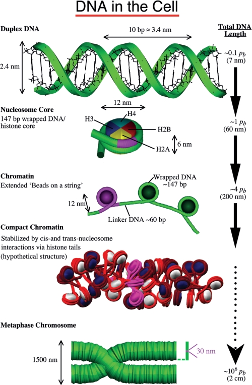
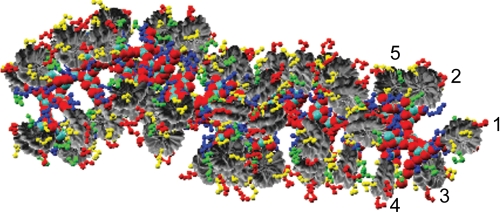
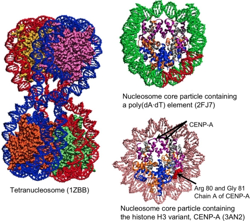
Understanding the structural organization of eukaryotic chromatin and its control of gene expression represents one of the most fundamental and open challenges in modern biology. Recent experimental advances have revealed important characteristics of chromatin in response to changes in external conditions and histone composition, such as the conformational complexity of linker DNA and histone tail domains upon compact folding of the fiber. In addition, modeling studies based on high-resolution nucleosome models have helped explain the conformational features of chromatin structural elements and their interactions in terms of chromatin fiber models. This minireview discusses recent progress and evidence supporting structural heterogeneity in chromatin fibers, reconciling apparently contradictory fiber models.


Figures

FIGURE 1.
FIGURE 1.
Schematic view of the many levels of DNA folding in the cell. On length scales much smaller than the persistence length (pb), DNA can be considered straight. In eukaryotic cells, DNA wraps around a core of histone proteins to form the chromatin fiber. The fiber is shown in both the extended view and a hypothetical compact zigzag view (the “30-nm fiber”) deduced from a modeling study (29). Chromosomes are made up of a dense chromatin fiber, shown here in the metaphase stage. For reference, we highlight in pink in all the DNA/protein views the hierarchical organizational unit preceding it. The length scale on the right indicates the level of compaction involved.
FIGURE 2.
FIGURE 2.
Detailed view of the heteromorphic chromatin model shown in Fig. 1, with rendering of the core histone tails to show the complex inter- and intranucleosome interactions. The first five nucleosomes are marked to indicate the different interaction types. Histone tails are colored yellow (H2A), red (H2B), blue (H3), and green (H4).
FIGURE 3.
FIGURE 3.
Examples of crystal structures for the tetranucleosome and nucleosomes. Shown are the tetranucleosome complex (25), a nucleosome core particle containing a poly(dA·dT) tract (red) (12), and a human centromeric nucleosome containing the centromere-specific histone H3 variant CENP-A (18). In single nucleosome particles, the histone proteins are colored by type (i.e. H3, purple; H4, silver; H2A, orange; H2B, blue). Arrows point to residues present in CENP-A but not in the canonical H3 histone.
FIGURE 4.
FIGURE 4.
Representative studies of chromatin structure providing foundations for the mesoscopic model of the 30-nm fiber. The models were selected among the many relevant works and are thus representative rather than complete. AFM, atomic force microscopy; SMFS, single-molecule force spectroscopy.
FIGURE 5.
FIGURE 5.
Chromatin organization: ideal and deduced models for relaxed chromatin and stretched chromatin fibers. A–C, solenoid, zigzag, and heteromorphic models, respectively. A, ideal model (parallel and perpendicular views) for a 48-core 209-bp one-start solenoid fiber with six nucleosomes/turn in which DNA linkers (DNA segments shown in red connecting nucleosomes) are bent and neighboring nucleosomes (i±1 interactions (int.)) are in the closest contact. B, ideal two-start zigzag model (parallel and perpendicular views) for a 48-core 209-bp fiber in which DNA linkers are straight and i±2 nucleosomes are in the closest contact. C, heteromorphic architecture predicted by modeling and Monte Carlo simulations of 48-core 209-bp arrays with LH at room temperature (293 K), 0.15 m NaCl, and low concentration of magnesium ions and confirmed by cross-linking experiments (29). DNA linkers are shown in red, alternate nucleosomes are shown in white and blue, and LHs are shown as turquoise spheres. The view parallel to the fiber axis (left) and two enlarged nucleosome triplets are shown. Both straight and bent DNA linkers occur. In all views, connecting DNA linkers and DNA wrapped around the nucleosomes are colored in red; odd and even nucleosomes are white and blue, respectively; and LHs are shown in turquoise. The close-ups of trinucleosomes show both intra- and internucleosome interactions. The core histone tails are colored yellow (H2A), red (H2B), blue (H3), and green (H4). D, effect of NRL (173, 209, and 218 bp) and LH on the structure of the chromatin fiber as predicted from Monte Carlo simulations of 48-core arrays at 0.15 m monovalent ions (72). The center images also show the individual histone tail beads. Color coding is as described above. E, effect of various dynamic LH binding mechanisms on the chromatin unfolding mechanism for 24-core 209-bp fibers as revealed from stretching simulations mimicking single-molecule pulling experiments at monovalent salt conditions of 0.15 m (43). Panel 1, resulting force extension curves for fibers with one LH rigidly fixed to each core (blue curve) versus LHs that bind/unbind dynamically (average concentration of 0.8 LH/core; red curve) with added divalent cations. Dynamic LH binding/unbinding dramatically decreases the fiber stiffness and the forces needed for unfolding with respect to fibers with fixed LH, improving the agreement with experiments (33) significantly. pN, piconewtons. Panel 2, images representing unfolding intermediates at different forces along the dynamic LH curve in panel 1. Intermediates reveal “superbead-on-a-string” structures in which compact clusters coexist with extended fiber regions. Panel 3, effect of fast and slow dynamic LH binding/unbinding during chromatin fiber unfolding without divalent ions (43). The slow-rebinding LH molecules cause a more dramatic softening effect than a pool of fast-rebinding LH, whereas fast LH rebinding promotes formation of superbead-on-a-string conformations with compact clusters. Together, fast- and slow-binding LH pools provide facile fiber unfolding through heteromorphic superbead conformations.

References

    1. Woodcock C. L., Dimitrov S. (2001) Higher order structure of chromatin and chromosomes. Curr. Opin. Genet. Dev. 11, 130–135 - PubMed
    1. Bancaud A., Huet S., Daigle N., Mozziconacci J., Beaudouin J., Ellenberg J. (2009) Molecular crowding affects diffusion and binding of nuclear proteins in heterochromatin and reveals the fractal organization of chromatin. EMBO J. 28, 3785–3798 - PMC - PubMed
    1. van Holde K. E. (1988) Chromatin, Springer-Verlag, New York
    1. Felsenfeld G., Groudine M. (2003) Controlling the double helix. Nature 421, 448–453 - PubMed
    1. Horn P. J., Peterson C. L. (2002) Molecular biology. Chromatin higher order folding–wrapping up transcription. Science 297, 1824–1827 - PubMed

Publication types

LinkOut - more resources