. 2012 Jan 27;287(5):3518-29.
doi: 10.1074/jbc.M111.317230. Epub 2011 Dec 7.

Nuclear import of the yeast hexokinase 2 protein requires α/β-importin-dependent pathway

Affiliations

Nuclear import of the yeast hexokinase 2 protein requires α/β-importin-dependent pathway

Rafael Peláez et al. J Biol Chem. .

Abstract

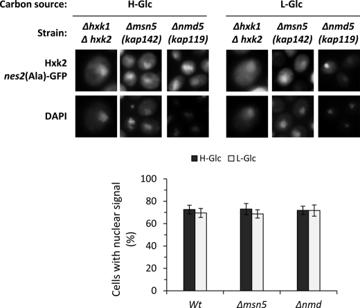
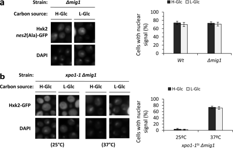
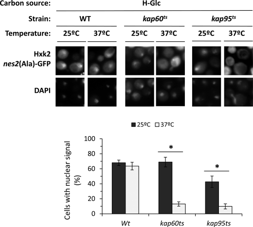
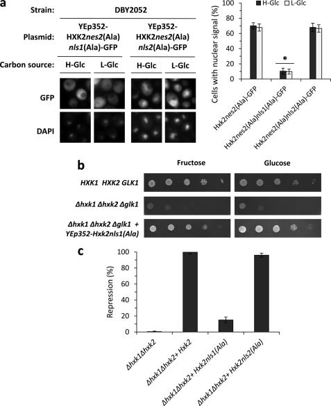
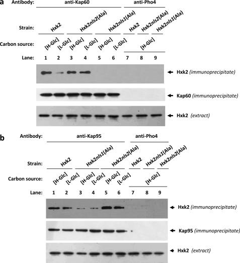
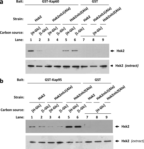
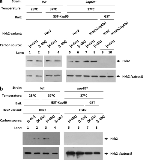
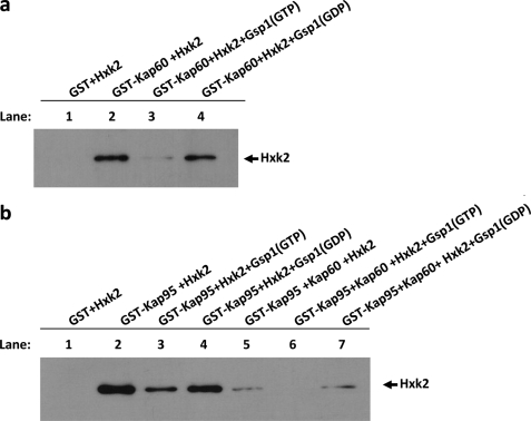
Hexokinase 2 (Hxk2) from Saccharomyces cerevisiae was one of the first metabolic enzymes described as a multifunctional protein. Hxk2 has a double subcellular localization and role, it functions as a glycolytic enzyme in the cytoplasm and as a regulator of gene transcription of several Mig1-regulated genes in the nucleus. However, the mechanism by which Hxk2 enters in the nucleus was unknown until now. Here, we report that the Hxk2 protein is an import substrate of the carriers α-importin (Kap60 in yeast) and β-importin (Kap95 in yeast). We also show that the Hxk2 nuclear import and the binding of Hxk2 with Kap60 are glucose-dependent and involve one lysine-rich nuclear localization sequence (NLS), located between lysine 6 and lysine 12. Moreover, Kap95 facilitates the recognition of the Hxk2 NLS1 motif by Kap60 and both importins are essential for Hxk2 nuclear import. It is also demonstrated that Hxk2 nuclear import and its binding to Kap95 and Kap60 depend on the Gsp1-GTP/GDP protein levels. Thus, our study uncovers Hxk2 as a new cargo for the α/β-importin pathway of S. cerevisiae.


Figures

FIGURE 1.
FIGURE 1.
Localization of Hxk2nes2(Ala) in Δhxk1Δhxk2, Δmsn5, and Δnmd5 yeast cells. The DBY2052, Y03694, and B0119B strains expressing Hxk2nes2(Ala)-GFP from plasmid YEp352-HXK2nes2(Ala)-GFP were grown in high-glucose synthetic medium (H-Glc) until an A600 nm of 1.0 was reached and then transferred to low-glucose synthetic medium (L-Glc) for 60 min. Cells were stained with DAPI and imaged for GFP and DAPI fluorescence. The nuclear localization of the Hxk2nes2(Ala)-GFP protein was determined in at least 100 cells per growth condition. No statistically significant differences were detected between mutants.
FIGURE 2.
FIGURE 2.
Localization of Hxk2nes2(Ala) in Δmig1 and xpo1–1Δmig1 yeast cells. a, the Δmig1 strain expressing Hxk2nes2(Ala)-GFP from plasmid YEp352-HXK2nes2(Ala)-GFP was grown in high-glucose synthetic medium (H-Glc) until an A600 nm of 1.0 was reached and then transferred to low-glucose synthetic medium (L-Glc) for 60 min. Cells were stained with DAPI and imaged for GFP and DAPI fluorescence. Nuclear localization of the Hxk2nes2(Ala)-GFP protein was determined in at least 100 cells per growth condition. Error bars represent mean ± S.D. for three independent experiments. b, the xpo1–1Δmig1 double mutant strain expressing Hxk2-GFP, from plasmid YEp352-HXK2/GFP, were grown in high-glucose synthetic medium (H-Glc) until an A600 nm of 1.0 was reached and then transferred to low-glucose synthetic medium (L-Glc). The cells were grown at 25 °C (permissive temperature) and then shifted to 37 °C (nonpermissive temperature) for 1 h. The localization of Hxk2-GFP was analyzed by fluorescence microscopy (GFP). Nuclear DNA was stained with DAPI. Nuclear localization of the Hxk2-GFP protein was determined in at least 100 cells per growth condition. No statistically significant differences were detected between mutants.
FIGURE 3.
FIGURE 3.
Nuclear import of Hxk2-GFP is inhibited in xpo1–1 cells at the nonpermissive temperature. The DBY2052, kap60ts, and kap95ts strains expressing Hxk2nes2(Ala)-GFP, from plasmid YEp352-HXK2nes2(Ala)/GFP, were grown in high-glucose synthetic medium (H-Glc) until an A600 nm of 1.0 was reached and then transferred to low-glucose synthetic medium (L-Glc). The cells were grown at 25 °C (permissive temperature) and then shifted to 37 °C (nonpermissive temperature) for 1 h. Cells were stained with DAPI and imaged for GFP and DAPI fluorescence. Nuclear localization of the Hxk2nes2(Ala)-GFP protein was determined in at least 100 cells per growth condition from 3 different experiments. *, statistically significant differences 25 versus 37 °C in Kaps mutants, p < 0.01.
FIGURE 4.
FIGURE 4.
Identification of Hxk2 NLSs. a, the DBY2052 (Δhxk1Δhxk2) mutant strain was transformed with plasmids YEp352-HXK2nes2(Ala)nls1(Ala)/GFP and YEp352-HXK2nes2(Ala)nls2(Ala)/GFP. Transformed cells were grown in high-glucose synthetic medium (H-Glc) until an A600 nm of 1.0 was reached and then transferred to low-glucose synthetic medium (L-Glc) for 60 min. The cells were visualized by fluorescence microscopy, DAPI staining revealed nuclear DNA. Nuclear localization of fluorescent reporter proteins was determined in at least 100 cells in three independent experiments. Mean ± S.D. are shown for at least three independent experiments. *, statistically significant differences between groups, p < 0.001. b, the THG1 (Δhxk1Δhxk2Δglk1) triple mutant strain was transformed with the YEp352-HXK2nls1(Ala) plasmid and grown overnight in synthetic media with galactose (2%) as carbon source (SGal ura). 5-Fold serial dilutions of a 1.0 A600 overnight culture were plated on SFru ura (fructose, 2%) medium and SD ura medium and photographed after 48 h. c, the Δhxk1Δhxk2 double mutant strain was transformed with YEp352, YEp352-HXK2, YEp352-HXK2nls1(Ala), and YEp352-HXK2nls2(Ala) plasmids. Transformed cells were grown in high-glucose synthetic medium until an A600 nm of 1.0 was reached. Invertase activity was assayed in whole cells. Values are the averages of results obtained on four independent experiments. *, statistically significant differences between groups, p < 0.01.
FIGURE 5.
FIGURE 5.
Interaction of Kap60 and Kap95 with Hxk2. In vivo co-immunoprecipitation of Kap60 (a) and Kap95 (b) with Hxk2, Hxk2nls1(Ala), and Hxk2nls2(Ala). The wild-type, FMY304, and FMY305 strains were grown in YEPD media until an A600 nm of 0.8 was reached and then shifted to high (H-Glc) and low (L-Glc) glucose conditions for 1 h. The cell extracts were immunoprecipitated with a polyclonal anti-Kap60 or anti-Kap95 antibodies (lanes 1–6), or a polyclonal antibody to Pho4 (lanes 7–9). Immunoprecipitates were separated by 12% SDS-PAGE, and co-precipitated Hxk2 variants were visualized on a Western blot with monoclonal anti-Hxk2 antibody. The level of immunoprecipitated Kap60 or Kap95 in the blotted samples was determined by using anti-Kap60 and anti-Kap95 antibodies, respectively. The level of Hxk2 present in the different extracts used in Fig. 5, a and b, was determined by Western blot using anti-Hxk2 antibody. The Western blots shown are representative of results obtained from four independent experiments.
FIGURE 6.
FIGURE 6.
GST pulldown assays of the interaction of Kap60 and Kap95 with Hxk2. The GST-Kap60 (a) and GST-Kap95 (b) fusion proteins were purified on glutathione-Sepharose columns. Equal amounts of GST-Kap60 and GST-Kap95 were incubated with cell extracts from wild-type, FMY304, and FMY305 strains. The yeast strains were grown in YEPD media until an A600 nm of 0.8 was reached and then shifted to low (L-Glc) glucose conditions for 1 h. After exhaustive washing the proteins were separated by 12% SDS-PAGE, and retained Hxk2 variants were visualized on a Western blot with polyclonal anti-Hxk2 antibody (lanes 1–6). For the control samples, GST protein was also incubated with the high-glucose (H-Glc) cell extracts, but no signals were detected (lanes 7–9). The level of Hxk2 present in the different extracts used in a and b was determined by Western blot using anti-Hxk2 antibody. The Western blots shown are representative of results obtained from four independent experiments.
FIGURE 7.
FIGURE 7.
GST pulldown assays of the interaction of Kap60 and Kap95 with Hxk2 in the absence of functional Kap95 and Kap60 proteins, respectively. The GST-Kap95 and GST-Kap60 fusion proteins were purified on glutathione-Sepharose columns. a, equal amounts of GST-Kap95 were incubated with cell extracts from wild-type, JCY1410 (kap60ts), and FMY306 (kap60tsHxk2nls1(Ala)) strains. b, equal amounts of GST-Kap60 were incubated with cell extracts from wild-type and JCY1407 (kap95ts) strains. The yeast strains were grown in YEPD media until an A600 nm of 0.8 was reached and then shifted to low (L-Glc) glucose conditions for 1 h. The wild-type strain was grown at 28 °C and the mutant cells were grown at 25 °C (permissive temperature) and then shifted to 37 °C (nonpermissive temperature) for 5 h. After exhaustive washing the proteins were separated by 12% SDS-PAGE, and the retained Hxk2 variants were visualized on a Western blot with polyclonal anti-Hxk2 antibody. For the control samples, GST protein was also incubated with the high-glucose (H-Glc) cell extracts, but no signals were detected. The level of Hxk2 present in the different extracts used in a and b was determined by Western blot using anti-Hxk2 antibody. The Western blots shown are representative of results obtained from four independent experiments.
FIGURE 8.
FIGURE 8.
The import Hxk2-Kap60-Kap95 complex formation. GST pulldown assays of the effect of Gsp1 on the Hxk2-Kap60 (a) and Hxk2-Kap95 (b) interaction stability. GST-Hxk2, GST-Gsp1, and GST-Kap60 fusion proteins were purified on glutathione-Sepharose columns and incubated with thrombin to isolate Hxk2, Gsp1, and Kap60 proteins, respectively. The Gsp1 protein was loaded with GTP to generate Gsp1(GTP), or with GDP to generate Gsp1(GDP). The Hxk2 protein was incubated with purified GST-Kap60 or GST-Kap95 bound to glutathione-Sepharose beads in the presence of Gsp1(GTP) and Gsp1(GDP) in the absence or presence of Kap60 purified protein (lanes 5–7). The beads were washed extensively. Co-precipitated proteins were resolved by 12% SDS-PAGE and visualized on a Western blot with polyclonal anti-Hxk2 antibody.

Similar articles

Cited by

References

    1. Radu A., Blobel G., Moore M. S. (1995) Identification of a protein complex that is required for nuclear protein import and mediates docking of import substrate to distinct nucleoporins. Proc. Natl. Acad. Sci. U.S.A. 92, 1769–1773 - PMC - PubMed
    1. Görlich D., Prehn S., Laskey R. A., Hartmann E. (1994) Isolation of a protein that is essential for the first step of nuclear protein import. Cell 79, 767–778 - PubMed
    1. Stade K., Ford C. S., Guthrie C., Weis K. (1997) Exportin 1 (Crm1p) is an essential nuclear export factor. Cell 90, 1041–1050 - PubMed
    1. Weis K. (2003) Regulating access to the genome. Nucleocytoplasmic transport throughout the cell cycle. Cell 112, 441–451 - PubMed
    1. Kalab P., Weis K., Heald R. (2002) Visualization of a Ran-GTP gradient in interphase and mitotic Xenopus egg extracts. Science 295, 2452–2456 - PubMed

Publication types

MeSH terms

LinkOut - more resources