. 2012 Feb 3;287(6):3850-8.
doi: 10.1074/jbc.M111.317404. Epub 2011 Dec 7.

Regulation of glycolytic enzyme phosphoglycerate mutase-1 by Sirt1 protein-mediated deacetylation

Affiliations

Regulation of glycolytic enzyme phosphoglycerate mutase-1 by Sirt1 protein-mediated deacetylation

William C Hallows et al. J Biol Chem. .

Abstract

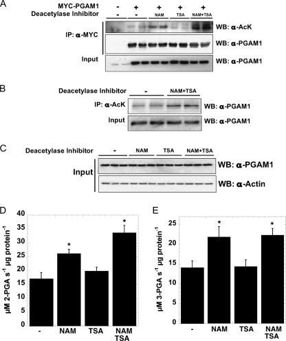
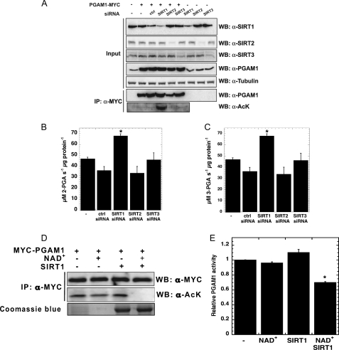
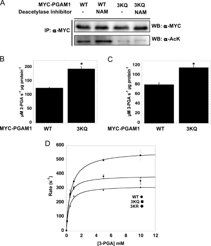
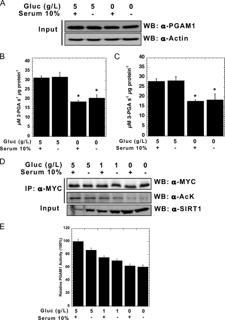
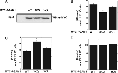
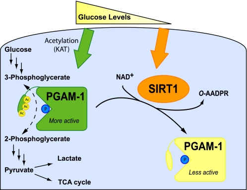
Emerging proteomic evidence suggests that acetylation of metabolic enzymes is a prevalent post-translational modification. In a few recent reports, acetylation down-regulated activity of specific enzymes in fatty acid oxidation, urea cycle, electron transport, and anti-oxidant pathways. Here, we reveal that the glycolytic enzyme phosphoglycerate mutase-1 (PGAM1) is negatively regulated by Sirt1, a member of the NAD(+)-dependent protein deacetylases. Acetylated PGAM1 displays enhanced activity, although Sirt1-mediated deacetylation reduces activity. Acetylation sites mapped to the C-terminal "cap," a region previously known to affect catalytic efficiency. Overexpression of a constitutively active variant (acetylated mimic) of PGAM1 stimulated flux through glycolysis. Under glucose restriction, Sirt1 levels dramatically increased, leading to PGAM1 deacetylation and attenuated activity. Previously, Sirt1 has been implicated in the adaptation from glucose to fat burning. This study (i) demonstrates that protein acetylation can stimulate metabolic enzymes, (ii) provides biochemical evidence that glycolysis is modulated by reversible acetylation, and (iii) demonstrates that PGAM1 deacetylation and activity are directly controlled by Sirt1.


Figures

FIGURE 1.
FIGURE 1.
PGAM1 is acetylated in vivo, and acetylation increases PGAM1 activity. A, transiently transfected PGAM1 is acetylated in vivo, when treated with sirtuin-specific inhibitors. MYC-PGAM1 was overexpressed in HEK293 cells and treated overnight with 5 mm NAM, 5 μm TSA, and 5 mm NAM, 5 μm TSA. MYC-PGAM1 was immunoprecipitated (IP) by anti-MYC conjugated to agarose at 4 °C for 4 h. These extracts were resolved by SDS-PAGE and detected by Western blotting (WB) with anti-acetyl-lysine (AcK) and anti-PGAM1 antibodies, respectively. Input refers to 1% of total lysate and was detected with anti-PGAM1 antibody. B, endogenous PGAM1 is acetylated in vivo. Endogenous PGAM1 is immunoprecipitated by anti-acetyl-lysine and protein G from HEK293 cells incubated overnight with 5 mm NAM, 5 μm TSA. These extracts (Input = 1% total cell lysate) were resolved by SDS-PAGE and detected by Western blotting with anti-PGAM1 antibody. C–E, soluble extracts of HEK293 cells incubated overnight with 5 mm NAM, 5 μm TSA, and 5 mm NAM, 5 μm TSA were resolved by SDS-PAGE and detected by Western blotting with anti-PGAM1 and anti-actin antibodies (C), respectively; activity assays measure PGAM1 activity. Input refers to 1% total cellular lysate. Forward PGAM reaction (D), coupled to Enolase, pyruvate kinase, and lactate dehydrogenase, utilized saturating 3-phosphoglycerate measuring absorbance at 340 nm. Reverse PGAM reaction (E), coupled to phosphoglycerate kinase and GAPDH, utilized saturating 3-phosphoglycerate measuring absorbance at 340 nm. Error bars represent S.E. (n = 3); *, p < 0.05 compared with untreated cells.
FIGURE 2.
FIGURE 2.
SIRT1 specifically deacetylates PGAM1. A, only SIRT1 knockdown significantly increased PGAM1-acetylated levels. HEK293 cells were transiently transfected with MYC-PGAM1 and siRNA to SIRT1–3. Soluble extracts were resolved by SDS-PAGE and detected through Western blot (WB), with anti-acetyl-lysine (AcK) and anti-PGAM1 antibodies. Input blots (1% total cellular extract) were detected with antibodies against PGAM1, SIRT1, SIRT2, SIRT3, and tubulin. B and C, activity was measured at 340 nm by PGAM1-coupled assay. D, acetylation of PGAM1 is decreased in vitro by SIRT1 deacetylation. Immunoprecipitants (IP) of MYC-PGAM1 from HEK293 cells transiently transfected with PGAM1 were incubated with the combination of NAD+ and SIRT1, and the reactions were resolved by SDS-PAGE and detected through Western blot, with anti-acetyl-lysine and anti-PGAM1 antibodies. E, activity was measured at 340 nm by PGAM1-coupled assay. Error bars represent S.E. (n = 3); *, p < 0.05 compared with control.
FIGURE 3.
FIGURE 3.
C-terminal tail of PGAM1 is acetylated. A, MYC-PGAM1 and MYC-PGAM1 3KQ mutant (K251Q, K253Q, and K254Q) were transiently transfected into HEK293 cells following 5 mm NAM treated for 16 h. Cell lysates were immunoprecipitated (IP) with anti-MYC antibody, resolved by SDS-PAGE, and then detected with Western blot (WB) analysis with anti-acetyl-lysine (AcK) and anti-MYC antibodies. B and C, activity was measured at 340 nm by the PGAM1-coupled assay. D, PGAM1 3KQ shows high catalytic activity against 2-phosphoglycerate. Catalytic amounts of PGAM1 ●, PGAM1 3KQ ■, and PGAM1 3KR ⧫ were incubated with the indicated amounts of 3-PGA, and activity was measured at 340 nm by PGAM1-coupled assay. Data from the coupled assay were fitted to the Michaelis-Menten equation. Error bars represent S.E. (n = 3). *, p < 0.05.
FIGURE 4.
FIGURE 4.
High glucose increases PGAM1 acetylation and activity in vivo. A, HEK293 cells were treated by 5 g/liter glucose (Gluc) ± FBS and 0 g/liter glucose ± FBS for 6 h. Endogenous PGAM1 activity assays were measured at 340 nm for both forward (B) and reverse reactions (C). Cell lysates were analyzed by SDS-PAGE and detected through Western blot (WB) with anti-PGAM1 and anti-actin antibodies. Input refers to 1% total cellular extract (A). D, MYC-PGAM1 was overexpressed in HEK293 cells following incubation for 6 h with 5 g/liter glucose ± FBS, 1 g/liter glucose ± FBS, and 0 g/liter glucose ± FBS. Cell lysates were resolved by SDS-PAGE, and Western blotting was performed with anti-SIRT1, anti-MYC, and anti-acetylated lysine antibodies. E, MYC-PGAM1 was immunoprecipitated (IP) with anti-MYC conjugated to agarose, and PGAM1 activity assay was measured. PGAM1 protein levels were normalized using quantification of the anti-MYC Western as in D. Input refers to 1% cellular extract. Error bars represent S.E. (n = 3); *, p < 0.05 compared with 5 g/liter glucose with serum.
FIGURE 5.
FIGURE 5.
Expression of PGAM1 mutant 3KQ (acetylated mimic) stimulates glycolytic flux. WT MYC-PGAM1, 3KQ, and 3KR were overexpressed in HEK293 cells. A, input blots (1% total lysate) were detected with anti-MYC antibody. B, glucose levels in HEK293 cells expressing PGAM1, PGAM1 3KQ, and PGAM1 3KR. C, lactate levels in HEK293 cells expressing PGAM1, PGAM1 3KQ, and PGAM1 3KR. D, alanine levels in HEK293 cells expressing PGAM1, PGAM1 3KQ, and PGAM1 3KR. Error bars represent S.E. (n = 3); *, p < 0.05.
FIGURE 6.
FIGURE 6.
Model of PGAM1 regulation by glucose availability and SIRT1-dependent reversible deacetylation. Under high glucose, acetylation of the C-terminal cap of PGAM1 stimulates catalysis. Under glucose restriction, SIRT1 levels increase, leading to deacetylation of PGAM1 and decreased enzymatic activity.

Similar articles

Cited by

References

    1. Smith B. C., Hallows W. C., Denu J. M. (2008) Mechanisms and molecular probes of sirtuins. Chem. Biol. 15, 1002–1013 - PMC - PubMed
    1. Hallows W. C., Lee S., Denu J. M. (2006) Sirtuins deacetylate and activate mammalian acetyl-CoA synthetases. Proc. Natl. Acad. Sci. U.S.A. 103, 10230–10235 - PMC - PubMed
    1. Schwer B., Bunkenborg J., Verdin R. O., Andersen J. S., Verdin E. (2006) Reversible lysine acetylation controls the activity of the mitochondrial enzyme acetyl-CoA synthetase 2. Proc. Natl. Acad. Sci. U.S.A. 103, 10224–10229 - PMC - PubMed
    1. Lin Y. Y., Lu J. Y., Zhang J., Walter W., Dang W., Wan J., Tao S. C., Qian J., Zhao Y., Boeke J. D., Berger S. L., Zhu H. (2009) Protein acetylation microarray reveals that NuA4 controls key metabolic target regulating gluconeogenesis. Cell 136, 1073–1084 - PMC - PubMed
    1. Hirschey M. D., Shimazu T., Goetzman E., Jing E., Schwer B., Lombard D. B., Grueter C. A., Harris C., Biddinger S., Ilkayeva O. R., Stevens R. D., Li Y., Saha A. K., Ruderman N. B., Bain J. R., Newgard C. B., Farese R. V., Jr., Alt F. W., Kahn C. R., Verdin E. (2010) SIRT3 regulates mitochondrial fatty acid oxidation by reversible enzyme deacetylation. Nature 464, 121–125 - PMC - PubMed