. 2012 Feb 17;287(8):5243-52.
doi: 10.1074/jbc.M111.306159. Epub 2011 Dec 7.

Multiple members of the UDP-GalNAc: polypeptide N-acetylgalactosaminyltransferase family are essential for viability in Drosophila

Affiliations

Multiple members of the UDP-GalNAc: polypeptide N-acetylgalactosaminyltransferase family are essential for viability in Drosophila

Duy T Tran et al. J Biol Chem. .

Abstract

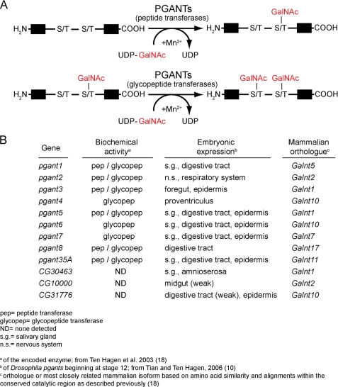
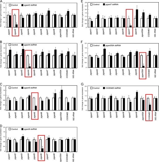
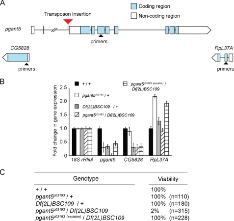
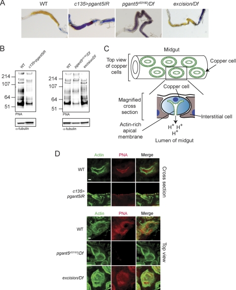
Mucin-type O-glycosylation represents a major form of post-translational modification that is conserved across most eukaryotic species. This type of glycosylation is initiated by a family of enzymes (GalNAc-Ts in mammals and PGANTs in Drosophila) whose members are expressed in distinct spatial and temporal patterns during development. Previous work from our group demonstrated that one member of this family is essential for viability and another member modulates extracellular matrix composition and integrin-mediated cell adhesion during development. To investigate whether other members of this family are essential, we employed RNA interference (RNAi) to each gene in vivo. Using this approach, we identified 4 additional pgant genes that are required for viability. Ubiquitous RNAi to pgant4, pgant5, pgant7, or the putative glycosyltransferase CG30463 resulted in lethality. Tissue-specific RNAi was also used to define the specific organ systems and tissues in which each essential family member is required. Interestingly, each essential pgant had a unique complement of tissues in which it was required. Additionally, certain tissues (mesoderm, digestive system, and tracheal system) required more than one pgant, suggesting unique functions for specific enzymes in these tissues. Expanding upon our RNAi results, we found that conventional mutations in pgant5 resulted in lethality and specific defects in specialized cells of the digestive tract, resulting in loss of proper digestive system acidification. In summary, our results highlight essential roles for O-glycosylation and specific members of the pgant family in many aspects of development and organogenesis.


Figures

FIGURE 1.
FIGURE 1.
Protein O-linked glycosylation. A, the enzymatic addition of N-acetylgalactosamine (GalNAc) to serine or threonine of protein substrates is catalyzed by the PGANT family of enzymes. The PGANT family is divided into 2 subgroups: peptide transferases, which catalyze the addition of GalNAc to unmodified substrates; and glycopeptide transferases, which only add GalNAc to previously glycosylated substrates. B, summarized is the pgant gene family from Drosophila, their embryonic expression patterns, the biochemical activity of the encoded enzymes, and the most closely related mammalian isoforms.
FIGURE 2.
FIGURE 2.
Quantitative PCR reveals specific decreases in gene expression of each pgant in progeny expressing dsRNA. Progeny expressing dsRNA to (A) pgant2, (B) pgant4, (C) pgant5, (D) pgant6, (E) pgant7, (F) pgant35A, and (G) CG30463 are shown. Total RNA was isolated and qPCR was performed for all pgant family members to verify specific knockdown of gene expression (red boxes). RNA levels were normalized to 18 S rRNA. Standard deviations are shown.
FIGURE 3.
FIGURE 3.
Transposon insertion mutation in pgant5 causes lethality and confirms pgant5 is an essential gene. A, the position of the transposon insertion in intron 2 of pgant5 is shown. Exons are represented as boxes and introns are represented as lines. Blue boxes are coding regions and white boxes are noncoding regions of pgant5. Flanking genes are also shown. Regions used to generate PCR primers are represented as triangles. B, real-time PCR analysis of pgant5 transcript levels using the primer pairs shown in A reveals a significant decrease in pgant5 gene expression in pgant5c03193/Df(2L)BSC109 relative to pgant5c03193/+ heterozygotes, Df(2L)BSC109/+ heterozygotes, pgant5c03193(excision)/Df(2L)BSC109, or wild type. RNA was normalized to 18S rRNA. C, loss of pgant5 results in lethality.
FIGURE 4.
FIGURE 4.
Loss of pgant5 results in loss of gut acidification and disruption of O-glycosylation in copper cells of the digestive tract. A, larval gut acidification as detected with bromophenol blue shows acidified midguts (yellow) in wild type and pgant5 transposon excision (excision/Df), but basic midguts (blue) in pgant5 mutants (pgant5c03193/Df) or larvae expressing dsRNA to pgant5 in the digestive system (c135>pgant5IR). B, Western blots of proteins from wild type, c135>pgant5IR, pgant5c03193/Df, and excision/Df third instar larval midguts probed with the lectin PNA to detect O-glycosylated proteins or with α-tubulin as a loading control. C, diagram of midgut and copper cell morphology. Shown is a diagram of a section of the midgut where copper cells stain for actin (green) in concentric circles. Below is a magnified cross-section of a copper cell, highlighting the unique invaginated actin-rich apical surface (green). D, copper cells from wild type, c135>pgant5IR, pgant5c03193/Df, and excision/Df third instar larval midguts were stained with PNA (red) and phalloidin/actin (green). In wild type copper cells, phalloidin staining labels actin-based microvilli unique to the apical region of copper cells. PNA-reactive O-glycoproteins are found along the apical and luminal regions of wild type copper cells (shown in upper panel cross-sections). All apical and luminal PNA staining is lost from c135>pgant5IR (upper panel) and pgant5c03193/Df copper cells (lower panel). Additionally, apical actin staining is irregular in size and shape in pgant5c03193/Df copper cells. PNA staining and gut acidification are restored upon excision of the transposon (excision/Df). Scale bars = 4 μm.

Similar articles

Cited by

References

    1. Hart G. W., Copeland R. J. (2010) Glycomics hits the big time. Cell 143, 672–676 - PMC - PubMed
    1. Liu L., Xu Y. X., Hirschberg C. B. (2010) The role of nucleotide sugar transporters in development of eukaryotes. Semin. Cell Dev. Biol. 21, 600–608 - PMC - PubMed
    1. Love D. C., Krause M. W., Hanover J. A. (2010) O-GlcNAc cycling. Emerging roles in development and epigenetics. Semin. Cell Dev. Biol. 21, 646–654 - PMC - PubMed
    1. Nakamura N., Lyalin D., Panin V. M. (2010) Protein O-mannosylation in animal development and physiology. From human disorders to Drosophila phenotypes. Semin. Cell Dev. Biol. 21, 622–630 - PMC - PubMed
    1. Schachter H. (2010) Mgat1-dependent N-glycans are essential for the normal development of both vertebrate and invertebrate metazoans. Semin. Cell Dev. Biol. 21, 609–615 - PubMed

Publication types

MeSH terms