

Leiomodin 1, a new serum response factor-dependent target gene expressed preferentially in differentiated smooth muscle cells
- PMID: 22157009
- PMCID: PMC3268406
- DOI: 10.1074/jbc.M111.302224
Leiomodin 1, a new serum response factor-dependent target gene expressed preferentially in differentiated smooth muscle cells
Abstract
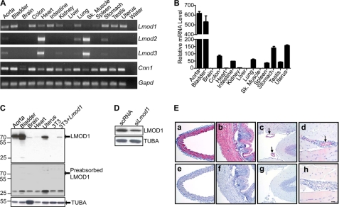
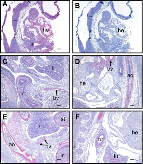
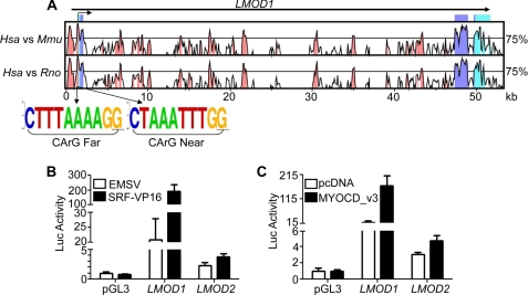
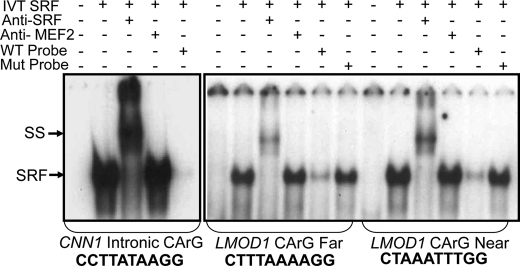
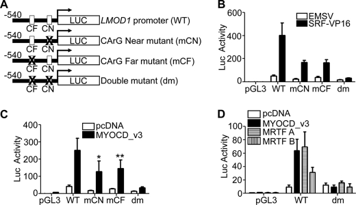
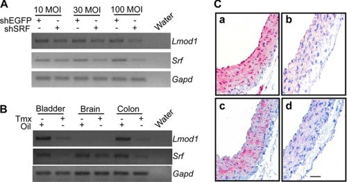
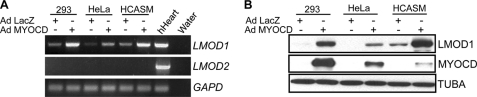
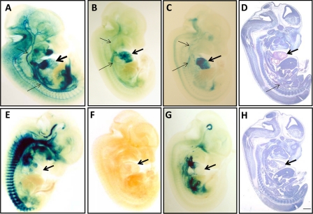
Smooth muscle cell (SMC) differentiation is defined largely by a number of cell-restricted genes governed directly by the serum response factor (SRF)/myocardin (MYOCD) transcriptional switch. Here, we describe a new SRF/MYOCD-dependent, SMC-restricted gene known as Leiomodin 1 (Lmod1). Conventional and quantitative RT-PCRs indicate that Lmod1 mRNA expression is enriched in SMC-containing tissues of the mouse, whereas its two paralogs, Lmod2 and Lmod3, exhibit abundant expression in skeletal and cardiac muscle with very low levels in SMC-containing tissues. Western blotting and immunostaining of various adult and embryonic mouse tissues further confirm SMC-specific expression of the LMOD1 protein. Comparative genomic analysis of the human LMOD1 and LMOD2 genes with their respective mouse and rat orthologs shows high conservation between the three exons and several noncoding sequences, including the immediate 5' promoter region. Two conserved CArG boxes are present in both the LMOD1 and LMOD2 promoter regions, although LMOD1 displays much higher promoter activity and is more responsive to SRF/MYOCD stimulation. Gel shift assays demonstrate clear binding between SRF and the two CArG boxes in human LMOD1. Although the CArG boxes in LMOD1 and LMOD2 are similar, only LMOD1 displays SRF or MYOCD-dependent activation. Transgenic mouse studies reveal wild type LMOD1 promoter activity in cardiac and vascular SMC. Such activity is abolished upon mutation of both CArG boxes. Collectively, these data demonstrate that Lmod1 is a new SMC-restricted SRF/MYOCD target gene.
Figures
Similar articles
-
The smooth muscle cell-restricted KCNMB1 ion channel subunit is a direct transcriptional target of serum response factor and myocardin.J Biol Chem. 2009 Nov 27;284(48):33671-82. doi: 10.1074/jbc.M109.050419. Epub 2009 Oct 1. J Biol Chem. 2009. PMID: 19801679 Free PMC article.
-
Contribution of serum response factor and myocardin to transcriptional regulation of smoothelins.Cardiovasc Res. 2006 Apr 1;70(1):136-45. doi: 10.1016/j.cardiores.2005.12.018. Epub 2006 Jan 31. Cardiovasc Res. 2006. PMID: 16451796
-
Smooth muscle expression of lipoma preferred partner is mediated by an alternative intronic promoter that is regulated by serum response factor/myocardin.Circ Res. 2008 Jul 3;103(1):61-9. doi: 10.1161/CIRCRESAHA.108.177436. Epub 2008 May 29. Circ Res. 2008. PMID: 18511849
-
Serum response factor: toggling between disparate programs of gene expression.J Mol Cell Cardiol. 2003 Jun;35(6):577-93. doi: 10.1016/s0022-2828(03)00110-x. J Mol Cell Cardiol. 2003. PMID: 12788374 Review.
-
Programming smooth muscle plasticity with chromatin dynamics.Circ Res. 2007 May 25;100(10):1428-41. doi: 10.1161/01.RES.0000266448.30370.a0. Circ Res. 2007. PMID: 17525382 Review.
Cited by
-
Transcriptomic Profiling of Skeletal Muscle Reveals Candidate Genes Influencing Muscle Growth and Associated Lipid Composition in Portuguese Local Pig Breeds.Animals (Basel). 2021 May 16;11(5):1423. doi: 10.3390/ani11051423. Animals (Basel). 2021. PMID: 34065673 Free PMC article.
-
Smooth muscle cell fate and plasticity in atherosclerosis.Cardiovasc Res. 2018 Mar 15;114(4):540-550. doi: 10.1093/cvr/cvy022. Cardiovasc Res. 2018. PMID: 29385543 Free PMC article. Review.
-
Putative Autoantigen Leiomodin-1 Is Expressed in the Human Brain and in the Membrane Fraction of Newly Formed Neurons.Pathogens. 2020 Dec 10;9(12):1036. doi: 10.3390/pathogens9121036. Pathogens. 2020. PMID: 33321732 Free PMC article.
-
Expression and promoter analysis of a highly restricted integrin alpha gene in vascular smooth muscle.Gene. 2013 Jan 15;513(1):82-9. doi: 10.1016/j.gene.2012.10.073. Epub 2012 Nov 8. Gene. 2013. PMID: 23142384 Free PMC article.
-
The N-terminal tropomyosin- and actin-binding sites are important for leiomodin 2's function.Mol Biol Cell. 2016 Aug 15;27(16):2565-75. doi: 10.1091/mbc.E16-03-0200. Epub 2016 Jun 15. Mol Biol Cell. 2016. PMID: 27307584 Free PMC article.
References
-
- Owens G. K., Kumar M. S., Wamhoff B. R. (2004) Molecular regulation of vascular smooth muscle cell differentiation in development and disease. Physiol. Rev. 84, 767–801 - PubMed
-
- Chow N., Bell R. D., Deane R., Streb J. W., Chen J., Brooks A., Van Nostrand W., Miano J. M., Zlokovic B. V. (2007) Serum response factor and myocardin mediate arterial hypercontractility and cerebral blood flow dysregulation in Alzheimer's phenotype. Proc. Natl. Acad. Sci. U.S.A. 104, 823–828 - PMC - PubMed
-
- Hirota J. A., Nguyen T. T., Schaafsma D., Sharma P., Tran T. (2009) Airway smooth muscle in asthma. Phenotype plasticity and function. Pulm. Pharmacol. Ther. 22, 370–378 - PubMed
-
- Norman C., Runswick M., Pollock R., Treisman R. (1988) Isolation and properties of cDNA clones encoding SRF, a transcription factor that binds to the c-fos serum response element. Cell 55, 989–1003 - PubMed
Publication types
MeSH terms
Substances
Grants and funding
LinkOut - more resources
Full Text Sources
Other Literature Sources
Molecular Biology Databases
Miscellaneous
