. 2012 Jan 27;287(5):3485-94.
doi: 10.1074/jbc.M111.296681. Epub 2011 Dec 6.

Lipin-1 phosphatidic phosphatase activity modulates phosphatidate levels to promote peroxisome proliferator-activated receptor γ (PPARγ) gene expression during adipogenesis

Affiliations

Lipin-1 phosphatidic phosphatase activity modulates phosphatidate levels to promote peroxisome proliferator-activated receptor γ (PPARγ) gene expression during adipogenesis

Peixiang Zhang et al. J Biol Chem. .

Abstract

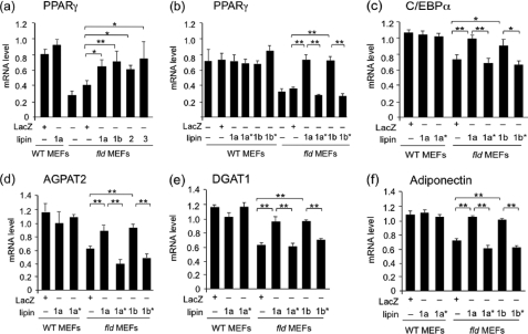
Adipose tissue plays a key role in metabolic homeostasis. Disruption of the Lpin1 gene encoding lipin-1 causes impaired adipose tissue development and function in rodents. Lipin-1 functions as a phosphatidate phosphatase (PAP) enzyme in the glycerol 3-phosphate pathway for triglyceride storage and as a transcriptional coactivator/corepressor for metabolic nuclear receptors. Previous studies established that lipin-1 is required at an early step in adipocyte differentiation for induction of the adipogenic gene transcription program, including the key regulator peroxisome proliferator-activated receptor γ (PPARγ). Here, we investigate the requirement of lipin-1 PAP versus coactivator function in the establishment of Pparg expression during adipocyte differentiation. We demonstrate that PAP activity supplied by lipin-1, lipin-2, or lipin-3, but not lipin-1 coactivator activity, can rescue Pparg gene expression and lipogenesis during adipogenesis in lipin-1-deficient preadipocytes. In adipose tissue from lipin-1-deficient mice, there is an accumulation of phosphatidate species containing a range of medium chain fatty acids and an activation of the MAPK/extracellular signal-related kinase (ERK) signaling pathway. Phosphatidate inhibits differentiation of cultured adipocytes, and this can be rescued by the expression of lipin-1 PAP activity or by inhibition of ERK signaling. These results emphasize the importance of lipid intermediates as choreographers of gene regulation during adipogenesis, and the results highlight a specific role for lipins as determinants of levels of a phosphatidic acid pool that influences Pparg expression.


Figures

FIGURE 1.
FIGURE 1.
Wild-type, but not PAP-deficient, lipins complement adipocyte differentiation in primary MEFs from fld mice. a, schematic diagram of lipin proteins, with evolutionarily conserved domains (N-LIP and C-LIP), nuclear localization signal (NLS), PAP enzyme (DIDGT), and coactivator motifs (LGHIL) indicated. The DIDGT motif was mutated to EIDGT in lipin-1a* (1a*) and lipin-1b* (1b*) to produce proteins that are PAP-deficient but retain ability to coactivate transcription (27, 35). Representative Western blot shows the protein levels of various lipin isoforms expressed in fld MEFs using adenovirus vectors and detected with anti-V5 antibody. b, lipin-1a and lipin-1a* exhibit similar transcriptional coactivator activity in MEFs. Cotransfection of fld MEFs with a luciferase reporter containing three tandem PPAR-response elements (PPRE) and expression vectors for PGC-1α, PPARγ, retinoid X receptor α, and lipin-1a or lipin-1a* led to the relative luciferase expression levels shown (n = 4 for each condition). c, overview of experimental design. Primary MEFs isolated from wild-type or fld mice were cultured to confluence and infected with adenoviruses expressing wild-type or PAP-deficient lipin proteins or LacZ as a negative control. Cells were subsequently stimulated to differentiate into adipocytes by treating with methylisobutylxanthine (MIX), dexamethasone (DEX), insulin, and rosiglitazone (TZD) for 2 days. Terminal adipocyte differentiation was promoted by continued culture in insulin and rosiglitazone from day 2 to day 6. d, representative Oil Red O staining of mature adipocytes (day 6 of differentiation) derived from wild-type (wt) or fld MEFs infected with adenoviruses expressing LacZ, wild-type lipin-1a, or PAP-deficient lipin-1a*. e, quantitation of cellular triglyceride content in MEFs at day 6 of adipocyte differentiation. Data represent the mean ± S.D. of triplicate determinations normalized to cellular protein. Statistically significant comparisons are identified with brackets; *, p < 0.05; **, p < 0.01.
FIGURE 2.
FIGURE 2.
Wild-type lipins, but not PAP-deficient coactivator-proficient lipins, complement the adipogenic gene transcription defect in primary MEFs from fld mice. qPCR was performed to determine PPARγ (a and b), C/EBPα (c), AGPAT2 (d), DGAT1 (e), and adiponectin (f) transcripts in wild-type or fld MEFs at post-differentiation day 6. Data are means ± S.D. of triplicate determinations normalized to 18 S RNA. *, p < 0.05; **, p < 0.01 for comparisons indicated.
FIGURE 3.
FIGURE 3.
Lipin-1 overcomes PA inhibition of PPARγ and C/EBPα gene expression during adipocyte differentiation. a, 3T3-F442A preadipocytes were treated with PA at the indicated concentrations and intracellular PAP levels measured as described under “Experimental Procedures.” b–d, 3T3-F442A preadipocytes were treated with insulin in the presence or absence of PA at the concentrations indicated for 2 days. qPCR was performed to determine expression levels of PPARγ (b), C/EBPα (c), and the apoptosis marker caspase-3 (d). Data are means ± S.D. of triplicate determinations normalized to TBP. *, p < 0.05; **, p < 0.01, versus no insulin treatment; #, p < 0.05; #, p < 0.01, versus insulin alone treatment. e, treatment of 3T3-F442A preadipocytes with dioleoylglycerol (DG) at the indicated concentrations did not influence Pparg expression levels. *, p < 0.05 compared with cells without insulin treatment. f and g, PA inhibition of adipogenic gene expression is reversed by expression of lipin-1a but not lipin-1a*. MEFs from wild-type mice were infected with adenovirus expressing lipin-1a, lipin-1a*, or LacZ and then stimulated to differentiate in the presence or absence of PA for 2 days. qPCR was performed to determine mRNA levels. Data are means ± S.D. of triplicate determinations normalized to TBP. *, p < 0.05; **, p < 0.01 for comparisons indicated.
FIGURE 4.
FIGURE 4.
Accumulation of phospholipid species in adipose tissue of fld mice. Electron spray ionization-mass spectrometric analysis of phospholipid species in inguinal adipose tissue of wild-type (WT) and fld mice revealed altered levels of PA (a), ceramides (b), sphingomyelin (SM), ceramide, hexosylceramide (HexCer), dihexosylceramide (DiHexCer), PC, lysophosphatidylcholine (LysoPC), and ether-linked phosphatidylcholine (ePC) (c). Right panels in a and b show molecular species of PA and ceramides indicated by total number of carbons and number of double bonds. (n = 6); *, p < 0.05; **, p < 0.01 versus WT.
FIGURE 5.
FIGURE 5.
Activation of ERK1/2 contributes to PA inhibition of adipocyte differentiation. a, adipose tissues from wild-type (WT) and fld mice were analyzed for the activation of ERK1/2 by Western blotting with specific antibodies recognizing ERK1/2 or the phosphorylated form (P-Erk1/2), and bands were quantitated by densitometry (n = 3). b, qPCR evaluation of the ERK1/2 effectors EGR-1 (also known as Krox-24) and c-Myc, and the ceramide-regulated genes caspase-9 and p21. c, inhibition of ERK1/2 rescues the PA inhibitory effect on PPARγ induction during 3T3-L1 adipocyte differentiation. Two-day postconfluent 3T3-L1 cells were stimulated with differentiation mixture in the presence or absence PA for 2 days. PPARγ mRNA levels were determined by quantitative RT-PCR and normalized to TBP. Data are means ± S.D. of triplicate determinations. *, p < 0.05 and **, p < 0.01 versus WT (in a and b) or for comparisons indicated (in c). DMI, dexamethasone, methylisobutylxanthine, insulin adipogenic mixture; PD, PD98059 MEK inhibitor.

References

    1. Huang-Doran I., Sleigh A., Rochford J. J., O'Rahilly S., Savage D. B. (2010) Lipodystrophy. Metabolic insights from a rare disorder. J. Endocrinol. 207, 245–255 - PubMed
    1. Lusis A. J., Attie A. D., Reue K. (2008) Metabolic syndrome. From epidemiology to systems biology. Nat. Rev. Genet. 9, 819–830 - PMC - PubMed
    1. Reue K., Phan J. (2006) Metabolic consequences of lipodystrophy in mouse models. Curr. Opin. Clin. Nutr. Metab. Care 9, 436–441 - PubMed
    1. Zalesin K. C., Franklin B. A., Miller W. M., Peterson E. D., McCullough P. A. (2011) Impact of obesity on cardiovascular disease. Med. Clin. North Am. 95, 919–937 - PubMed
    1. Lowe C. E., O'Rahilly S., Rochford J. J. (2011) Adipogenesis at a glance. J. Cell Sci. 124, 2681–2686 - PubMed

Publication types

MeSH terms