Method for expressing clinical and statistical significance of ocular and corneal wave front error aberrations
- PMID: 22157570
- PMCID: PMC3276750
- DOI: 10.1097/ICO.0b013e318221ce7d
Method for expressing clinical and statistical significance of ocular and corneal wave front error aberrations
Abstract
Purpose: The significance of ocular or corneal aberrations may be subject to misinterpretation whenever eyes with different pupil sizes or the application of different Zernike expansion orders are compared. A method is shown that uses simple mathematical interpolation techniques based on normal data to rapidly determine the clinical significance of aberrations, without concern for pupil and expansion order.
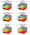
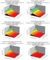
Methods: Corneal topography maps (TOMEY, Inc, Nagoya, Japan) from 30 normal corneas were collected, and the corneal wave front error was analyzed by Zernike polynomial decomposition into specific aberration types for pupil diameters of 3, 5, 7, and 10 mm and Zernike expansion orders of 6, 8, 10, and 12. Using this 4 × 4 matrix of pupil sizes and fitting orders, the best-fitting 3-dimensional functions were determined for the mean and standard deviation of the root-mean-square error for specific aberrations. The functions were encoded into a software application to determine the significance of data acquired from nonnormal cases.
Results: The best-fitting functions for 6 types of aberrations were determined: defocus, astigmatism, prism, coma, spherical aberration, and all higher-order aberrations. A clinical screening method of color coding the significance of aberrations in normal, postoperative laser in situ keratomileusis, and keratoconus cases having different pupil sizes and different expansion orders is demonstrated.
Conclusions: A method to calibrate wave front aberrometry devices using a standard sample of normal cases was devised. This method could be potentially useful in clinical studies involving patients with uncontrolled pupil sizes or in studies that compare data from aberrometers that use different Zernike fitting-order algorithms.
Conflict of interest statement
The author has no commercial or financial interest in the products or methods described in this manuscript.
Figures


Similar articles
-
LASIK-induced aberrations: comparing corneal and whole-eye measurements.Optom Vis Sci. 2015 Apr;92(4):447-55. doi: 10.1097/OPX.0000000000000557. Optom Vis Sci. 2015. PMID: 25785529
-
Shifting of the line of sight in keratoconus measured by a hartmann-shack sensor.Ophthalmology. 2010 Jan;117(1):41-8. doi: 10.1016/j.ophtha.2009.06.039. Epub 2009 Nov 6. Ophthalmology. 2010. PMID: 19896193
-
Goodness-of-prediction of Zernike polynomial fitting to corneal surfaces.J Cataract Refract Surg. 2005 Dec;31(12):2350-5. doi: 10.1016/j.jcrs.2005.05.025. J Cataract Refract Surg. 2005. PMID: 16473230
-
[Monochromatic aberration in accommodation. Dynamic wavefront analysis].Ophthalmologe. 2011 Jun;108(6):553-60. doi: 10.1007/s00347-011-2336-7. Ophthalmologe. 2011. PMID: 21695608 Review. German.
-
[Quantitative assessment of quality of vision].Nippon Ganka Gakkai Zasshi. 2004 Dec;108(12):770-807; discussion 808. Nippon Ganka Gakkai Zasshi. 2004. PMID: 15656087 Review. Japanese.
Cited by
-
Excimer Laser Surgery: Biometrical Iris Eye Recognition with Cyclorotational Control Eye Tracker System.Sensors (Basel). 2017 May 25;17(6):1211. doi: 10.3390/s17061211. Sensors (Basel). 2017. PMID: 28587100 Free PMC article.
-
Wavefront aberration changes caused by a gradient of increasing accommodation stimuli.Eye (Lond). 2015 Jan;29(1):115-21. doi: 10.1038/eye.2014.244. Epub 2014 Oct 24. Eye (Lond). 2015. PMID: 25341432 Free PMC article.
-
Clinical applications of wavefront refraction.Optom Vis Sci. 2014 Oct;91(10):1278-86. doi: 10.1097/OPX.0000000000000377. Optom Vis Sci. 2014. PMID: 25216319 Free PMC article. Review.
References
-
- Mrochen M, Kaemmerer M, Seiler T. Clinical results of wavefront-guided laser in-situ keratomileusis 3 months after surgery. J Cataract Refract Surg. 2001;27:201–207. - PubMed
-
- Marcos S, Barbero S, Llorente L, Merayo-Lloves J. Optical response to LASIK surgery for myopia from total and corneal aberration measurements. Invest Ophthalmol Vis Sci. 2001;42:3349–3356. - PubMed
-
- Zhou C, Chai X, Yuan L, He Y, Jin M, Ren Q. Corneal higher-order aberrations after customized aspheric ablation and conventional ablation for myopic correction. Curr Eye Res. 2007;32:431–438. - PubMed
-
- Scharma M, Wachler BS, Chan CC. Higher order aberrations and relative risk of symptoms after LASIK. J Refract Surg. 2007;23:252–256. - PubMed
-
- de Castro LE, Sandoval HP, Bartholomew LR, Vroman DT, Solomon KD. High-order aberrations and preoperative associated factors. Acta Ophthalmologica Scan. 2007;85:106–110. - PubMed
Publication types
MeSH terms
Grants and funding
LinkOut - more resources
Full Text Sources

