Review
. 2012 Mar;81(5):434-41.
doi: 10.1038/ki.2011.399. Epub 2011 Dec 7.

Membranoproliferative glomerulonephritis and C3 glomerulopathy: resolving the confusion

Affiliations
Review

Membranoproliferative glomerulonephritis and C3 glomerulopathy: resolving the confusion

Sanjeev Sethi et al. Kidney Int. 2012 Mar.

Abstract

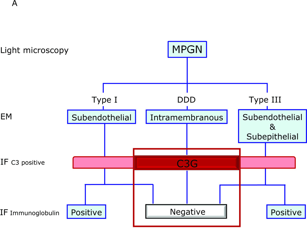
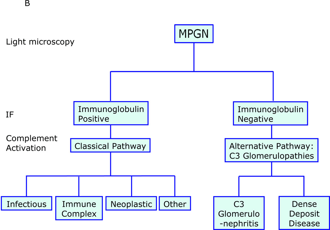
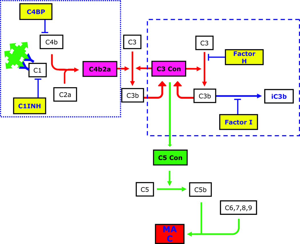
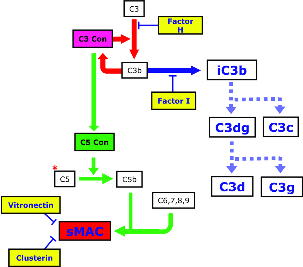
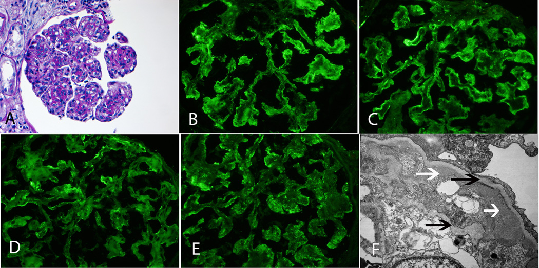
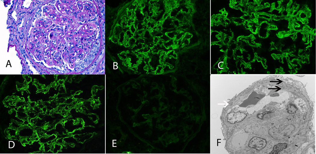
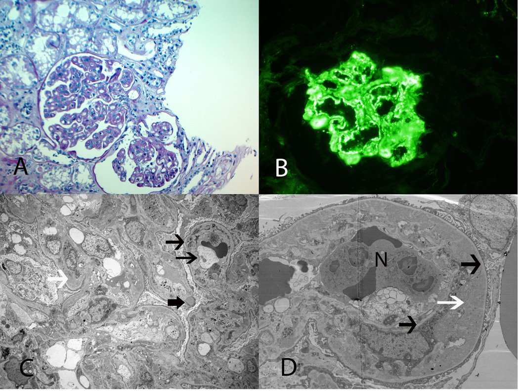
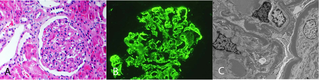
Membranoproliferative glomerulonephritis (MPGN) denotes a general pattern of glomerular injury that is easily recognized by light microscopy. With additional studies, MPGN subgrouping is possible. For example, electron microscopy resolves differences in electron-dense deposition that are classically referred to as MPGN type I (MPGN I), MPGN II, and MPGN III, while immunofluorescence typically detects immunoglobulins in MPGN I and MPGN III but not in MPGN II. All three MPGN types stain positive for complement component 3 (C3). Subgrouping has led to unnecessary confusion, primarily because immunoglobulin-negative MPGN I and MPGN III are more common than once recognized. Together with MPGN II, which is now called dense deposit disease, immunoglobulin-negative, C3-positive glomerular diseases fall under the umbrella of C3 glomerulopathies (C3G). The evaluation of immunoglobulin-positive MPGN should focus on identifying the underlying trigger driving the chronic antigenemia or circulating immune complexes in order to begin disease-specific treatment. The evaluation of C3G, in contrast, should focus on the complement cascade, as dysregulation of the alternative pathway and terminal complement cascade underlies pathogenesis. Although there are no disease-specific treatments currently available for C3G, a better understanding of their pathogenesis would set the stage for the possible use of anti-complement drugs.


Figures

Figure 1
Figure 1
A: MPGN-based and C3G-based classifications of glomerular disease overlap and are confusing because these two classifications are driven by different starting points - findings on EM for MPGN and on IF for C3G. As a general rule, MPGN I and MPGN III are immune-complex diseases characterized by subendothelial or subendothelial/subepithelial densities resolved by EM, however examples of immunoglobulin-negative ‘MPGN I’ and ‘MPGN III’ have been recognized for decades. These types of pathology fall under the umbrella of C3G and are often called C3 Glomerulonephritis (C3-GN). MPGN II or DDD (the preferred name) is another type of C3G. B: A simpler classification is driven by findings on IF, and classifies MPGN as immunoglobulin positive or negative. The terms MPGNI and MPGNIII are not used, thereby avoiding unnecessary confusion. Immunoglobulin-positive MPGN suggests CP activation and a concerted effort should be made to identify the underlying cause of antigenemia. Immunoglobulin-negative, C3-positive MPGN is due to dysregulation of the AP and TCC. Depending of the relative degree of dysregulation, the EM picture can resemble DDD or C3GN. Non-MPGN C3 Glomerulopathies are also seen (see Table 2, Light Microscopy).
Figure 1
Figure 1
A: MPGN-based and C3G-based classifications of glomerular disease overlap and are confusing because these two classifications are driven by different starting points - findings on EM for MPGN and on IF for C3G. As a general rule, MPGN I and MPGN III are immune-complex diseases characterized by subendothelial or subendothelial/subepithelial densities resolved by EM, however examples of immunoglobulin-negative ‘MPGN I’ and ‘MPGN III’ have been recognized for decades. These types of pathology fall under the umbrella of C3G and are often called C3 Glomerulonephritis (C3-GN). MPGN II or DDD (the preferred name) is another type of C3G. B: A simpler classification is driven by findings on IF, and classifies MPGN as immunoglobulin positive or negative. The terms MPGNI and MPGNIII are not used, thereby avoiding unnecessary confusion. Immunoglobulin-positive MPGN suggests CP activation and a concerted effort should be made to identify the underlying cause of antigenemia. Immunoglobulin-negative, C3-positive MPGN is due to dysregulation of the AP and TCC. Depending of the relative degree of dysregulation, the EM picture can resemble DDD or C3GN. Non-MPGN C3 Glomerulopathies are also seen (see Table 2, Light Microscopy).
Figure 2
Figure 2
The complement cascade is initiated by the CP (dotted rectangle), AP (dashed rectangle) or LP (not shown). The principle trigger of the CP, immunoglobulin-complexed antigen, is the driving force for C3 deposition in immunoglobulin-positive MPGN (see Figure 1B). Two or more adjacent IgG antibodies provide the structural framework for activation of C1, which cleaves C2 and C4 to generate C2a and C4b, respectively. These two proteins form the CP C3 convertase. The AP is constitutively active, a process that is referred to as ‘tick over’. Binding of CFB, CFD and properdin to hydrolyzed C3 or to C3b leads to formation of the AP C3 convertase C3bBb. This process also generates C3a, a potent anaphylatoxin. When an additional C3b molecule associates with C3 convertase, C5 convertase is formed, initiating the TCC and leading to the generation of MAC. Fluid-phase regulators of the CP include C4BP and C1INH, and of the AP, CFH and CFI (shown in yellow rectangles).
Figure 3
Figure 3
C3G is caused by dysregulation of the AP and TCC. The prototypic example is DDD, in which there must be fluid-phase dysregulation of the C3 convertase, often with dysregulation of the C5 convertase. Dysregulation of the C3 convertase results in increased serum levels of CFI-generated iC3b, which accumulates in the GBM as the characteristic sausage-shaped dense deposits. C3b breakdown products are also detectable in plasma as C3c and C3d. Some patients with C3G also have elevated serum levels of sMAC. Why dysregulation of the AP and TCC leads to DDD in some patients and to C3GN in others is NOT understood at this time. However genetic evidence suggests that a contributing factor may be the relative degree of dysregulation of the C3 and C5 convertases. For example, in the family described by Martínez-Barricarte and colleagues, there is dysregulation of C3 convertase but not C5 convertase. Clarifying the differences between DDD and C3GN is likely to be germane to the treatment of these diseases as the availability of anti-complement therapies is currently limited to eculizumab, an anti-C5 monoclonal antibody (shown as a red *) that prevents cleavage of C5 to C5a and C5b, thereby preventing propagation of the TCC.
Figure 4
Figure 4
MPGN secondary to Hepatitis C. A: LM shows MPGN with mesangial and endocapillary proliferation and thickened capillary walls with double contour formation (PAS ×20). B, C, D, E: IF microscopy shows mesangial and capillary wall staining for IgM, C3, kappa and lambda light chains (×40). F: EM shows subendothelial deposits (white arrows) with double contour formation (black arrows) (×13500).
Figure 5
Figure 5
MPGN secondary to monoclonal immunoglobulins. A: LM shows an MPGN with mesangial and endocapillary proliferation and thickened capillary walls with double contour formation. A small cellular crescent is also noted (PAS ×40). B, C, D, E: IF microscopy shows mesangial and capillary wall staining for IgG, C3 and kappa light chains but is negative for lambda light chains (×40). F: EM shows small subendothelial deposits (white arrow) with double contour formation (black arrows) (×3400).
Figure 6
Figure 6
C3GN. A: LM shows MPGN with thickened capillary walls and double contour formation (PAS ×20). B: IF microscopy shows strong capillary wall staining for C3 (×40). C, D: EM shows large subendothelial deposits (white arrows), occasional subepithelial deposit (thick black arrow) with double contour formation (thin black arrows) (N, neutrophil) (C ×1650, D ×4200).
Figure 7
Figure 7
Dense Deposit Disease. A: LM shows MPGN (H and E, ×20). B: IF microscopy showing mesangial and capillary wall staining for C3. C: EM shows abundant dense deposits along the glomerular basement membranes and mesangium (×3400).

Comment in

  • The many faces of C3 glomerulopathy.
    Meleg-Smith S. Meleg-Smith S. Kidney Int. 2012 Sep;82(5):611; author reply 611. doi: 10.1038/ki.2012.220. Kidney Int. 2012. PMID: 22892860 No abstract available.

References

    1. Anders D, Thoenes W. Basement membrane changes in membranoproliferative glomerulonephritis: a light and electron microscopic study. Virchows Arch Pathol Anat. 1975;369:87–109. - PubMed
    1. Habib R, Kleinknecht C, Gubler MC, Levy M. Idiopathic membranoproliferative glomerulonephritis in children. Report of 105 cases. Clin Nephrol. 1973;1:194–214. - PubMed
    1. Jackson EC, McAdams AJ, Strife CF, et al. Differences between membranoproliferative glomerulonephritis types I and III in clinical presentation, glomerular morphology and complement pertubation. Am J Kidney Dis. 1987;9:115–120. - PubMed
    1. Strife CF, McEnery PT, McAdams AJ, et al. Membranoproliferative glomerulonephritis with disruption of the glomerular basement membrane. Clin Nephrol. 1977;7:65–72. - PubMed
    1. Levy M, Gubler M-C, Sich M, et al. Immunopathology of membranoproliferative glomerulonephritis with subendothelial deposits (Type I MPGN) Clin Immunol Immunopathol. 1978;10:477–492. - PubMed

Publication types

MeSH terms