. 2011 Dec 11;18(1):148-52.
doi: 10.1038/nm.2574.

Focal adhesion kinase links mechanical force to skin fibrosis via inflammatory signaling

Affiliations

Focal adhesion kinase links mechanical force to skin fibrosis via inflammatory signaling

Victor W Wong et al. Nat Med. .

Abstract

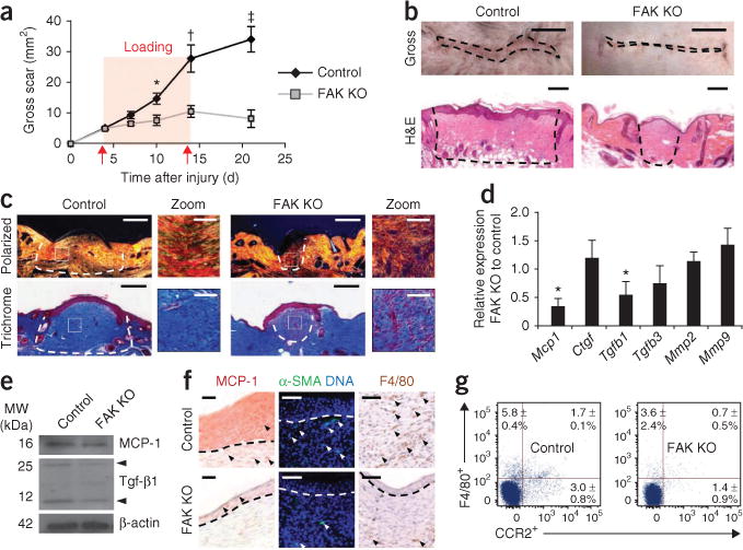
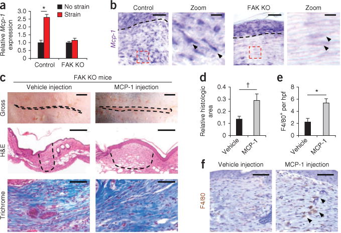
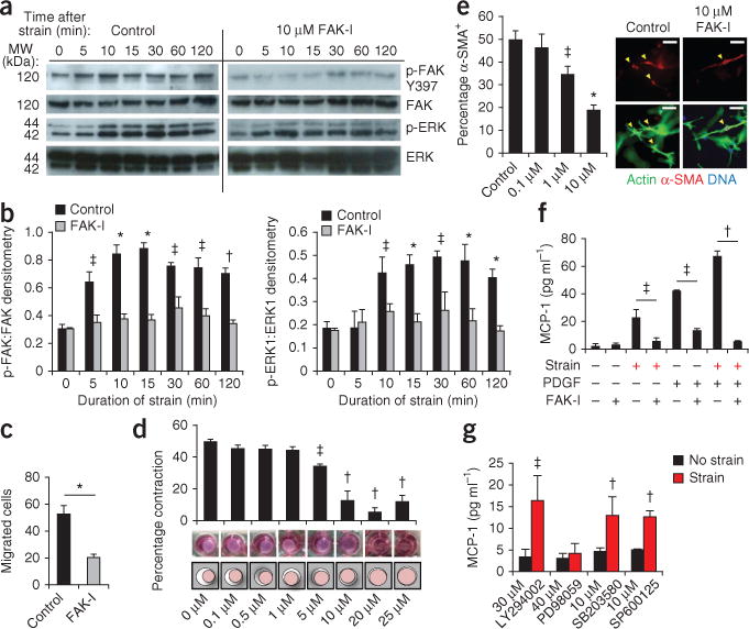
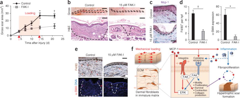
Exuberant fibroproliferation is a common complication after injury for reasons that are not well understood. One key component of wound repair that is often overlooked is mechanical force, which regulates cell-matrix interactions through intracellular focal adhesion components, including focal adhesion kinase (FAK). Here we report that FAK is activated after cutaneous injury and that this process is potentiated by mechanical loading. Fibroblast-specific FAK knockout mice have substantially less inflammation and fibrosis than control mice in a model of hypertrophic scar formation. We show that FAK acts through extracellular-related kinase (ERK) to mechanically trigger the secretion of monocyte chemoattractant protein-1 (MCP-1, also known as CCL2), a potent chemokine that is linked to human fibrotic disorders. Similarly, MCP-1 knockout mice form minimal scars, indicating that inflammatory chemokine pathways are a major mechanism by which FAK mechanotransduction induces fibrosis. Small-molecule inhibition of FAK blocks these effects in human cells and reduces scar formation in vivo through attenuated MCP-1 signaling and inflammatory cell recruitment. These findings collectively indicate that physical force regulates fibrosis through inflammatory FAK-ERK-MCP-1 pathways and that molecular strategies targeting FAK can effectively uncouple mechanical force from pathologic scar formation.


Conflict of interest statement

COMPETING FINANCIAL INTERESTS

The authors declare no competing financial interests.

Figures

Figure 1
Figure 1
HTS model. (a) Surface scar formation. n = 6. (b) Images of scars at 10 d after injury. Scale bars, top, 0.5 cm; bottom, 200 μm. (c) Polarized light and trichrome-stained images. n = 6. Scale bars, 200 μm; zoom scale bars, 50 μm. (d) Quantitative RT-PCR (qRT-PCR) analysis of wound cytokines. n = 9. (e) Scar cytokine densitometry. Arrowheads point to the monomer and dimer forms of Tgf-β1. n = 3. (f) Immunolocalization of MCP-1, α-SMA+ cells and F4/80+ macrophages. Scale bars, left column, 20 μm; middle and right columns, 50 μm. n = 6. (g) F4/80+ and CCR2+ flow cytometry. Quadrant values represent the percentage of total scar cells. n = 4. Values are means ± s.e.m. *P < 0.05, †P < 0.01, ‡P < 0.001. The dashed lines outline the scar.
Figure 2
Figure 2
Fibroblast-specific MCP-1 pathways. (a) Mcp-1 transcription in strained fibroblasts. n = 6. (b) Mcp-1 in situ hybridization at day 10 after injury (Mcp-1 transcripts are stained purple). Arrowheads point to spindle-shaped fibroblasts in the hpf (dashed red boxes). The dashed black lines indicate the basement membrane. Scale bars for the zoom images, 10 μm. (c) Analysis of fibrosis after intradermal injection of recombinant mouse MCP-1. The dashed lines outline the scar. Scale bars, top row, 0.25 cm; middle row, 200 μm. (d) Scar area at day 10 after injury. n = 6. (e,f) Quantification (e) and images (f) of F4/80+ macrophages in FAK knockout scars treated with vehicle or MCP-1. The arrowheads point to the macrophages. Values are means ± s.e.m. Scale bars, 50 μm unless otherwise noted. *P < 0.001, †P < 0.05.
Figure 3
Figure 3
FAK-mediated mechanoresponsive pathways in human fibroblasts. (a,b) Representative immunoblots and quantification of static strain-induced FAK and ERK activation in untreated human fibroblasts (Control) and those treated with the FAK inhibitor (FAK-I) PF573228. n = 3. (c) Fibroblast motility in a scratch migration assay. n = 6. (d,e) Fibroblast contraction (d) and α-SMA+ expression (arrowheads) (e) in three dimensional collagen lattices. Concentrations of PF573228 are indicated along the x-axes of the bar graphs. n = 3. Scale bars, 50 μm. (f) Synergistic (strain plus 10 ng ml−1 platelet-derived growth factor) induction of MCP-1 secretion. n = 4. (g) Strain-induced MCP-1 secretion with small-molecule inhibition of FAK (PF573228), Akt (LY294002), ERK (PD98059), p38 (SB203580) or JNK (SP600125). n = 4. Values are means ± s.e.m. *P < 0.001, †P < 0.01, ‡P < 0.05.
Figure 4
Figure 4
Intradermal treatment with PF573228. (a) Surface scar formation with treatment with 15 μM PF573228. n = 6. (b) Images of scars at day 10 after injury. The dashed lines indicate scar area. Scale bars, top row, 0.25; micrographs, 100 μm. n = 6. (c) Mcp-1 immunolocalization (purple color). The dashed lines indicate the basement membrane. Scale bars, 50 μm. (d,e) Quantification (d) and micrographs (e) of F4/80+ macrophages and α-SMA expression. n = 6. Scale bars, top row, 50 μm; bottom row, 100 μm. Values are means ± s.e.m. *P < 0.01, †P < 0.05. (f) Schematic of the proposed vicious cycle of hypertrophic scarring driven by mechanical activation of local and systemic fibroproliferative pathways through fibroblast FAK.

References

    1. Gurtner GC, Werner S, Barrandon Y, Longaker MT. Wound repair and regeneration. Nature. 2008;453:314–321. - PubMed
    1. Parsons JT. Focal adhesion kinase: the first ten years. J Cell Sci. 2003;116:1409–1416. - PubMed
    1. Ferreira AM, et al. Diminished induction of skin fibrosis in mice with MCP-1 deficiency. J Invest Dermatol. 2006;126:1900–1908. - PubMed
    1. Wynn TA. Cellular and molecular mechanisms of fibrosis. J Pathol. 2008;214:199–210. - PMC - PubMed
    1. Wynn TA. Common and unique mechanisms regulate fibrosis in various fibroproliferative diseases. J Clin Invest. 2007;117:524–529. - PMC - PubMed

Publication types

MeSH terms

Substances

Associated data