. 2012 Feb 24;287(9):6604-14.
doi: 10.1074/jbc.M111.292664. Epub 2011 Dec 8.

BRCA2 protein deficiency exaggerates doxorubicin-induced cardiomyocyte apoptosis and cardiac failure

Affiliations

BRCA2 protein deficiency exaggerates doxorubicin-induced cardiomyocyte apoptosis and cardiac failure

Krishna K Singh et al. J Biol Chem. .

Abstract

The tumor suppressor breast cancer susceptibility gene 2 (BRCA2) plays an important role in the repair of DNA damage, and loss of BRCA2 predisposes carriers to breast and ovarian cancers. Doxorubicin (DOX) remains the cornerstone of chemotherapy in such individuals. However, it is often associated with cardiac failure, which once manifests carries a poor prognosis. Because BRCA2 regulates genome-wide stability and facilitates DNA damage repair, we hypothesized that loss of BRCA2 may increase susceptibility to DOX-induced cardiac failure. To this aim, we generated cardiomyocyte-specific BRCA2 knock-out (CM-BRCA2(-/-)) mice using the Cre-loxP technology and evaluated their basal and post-DOX treatment phenotypes. Although CM-BRCA2(-/-) mice exhibited no basal cardiac phenotype, DOX treatment resulted in markedly greater cardiac dysfunction and mortality in CM-BRCA2(-/-) mice compared with control mice. Apoptosis in left ventricular (LV) sections from CM-BRCA2(-/-) mice compared with that in corresponding sections from wild-type (WT) littermate controls was also significantly enhanced after DOX treatment. Microscopic examination of LV sections from DOX-treated CM-BRCA2(-/-) mice revealed a greater number of DNA double-stranded breaks and the absence of RAD51 focus formation, an essential marker of double-stranded break repair. The levels of p53 and the p53-related proapoptotic proteins p53-up-regulated modulator of apoptosis (PUMA) and Bax were significantly increased in samples from CM-BRCA2(-/-) mice. This corresponded with increased Bax to Bcl-2 ratios and elevated cytochrome c release in the LV sections of DOX-treated CM-BRCA2(-/-) mice. Taken together, these data suggest a critical and previously unrecognized role of BRCA2 as a gatekeeper of DOX-induced cardiomyocyte apoptosis and susceptibility to overt cardiac failure. Pharmacogenomic studies evaluating cardiac function in BRCA2 mutation carriers treated with doxorubicin are encouraged.


Figures

FIGURE 1.
FIGURE 1.
Generation of cardiomyocyte-specific BRCA2 knock-out (CM-BRCA2−/−) mice. A, breeding strategy for the generation of CM-BRCA2−/− mice using the Cre-loxP technology. B, schematic representation of the Cre-mediated deletion of exon 11 of the BRCA2 gene. Arrows denote the binding sites of the primers (12/13 and 14/15) used for genotypic identification. C, representative PCR products of gDNA isolated from the ear notches of mice using primer pairs 12/13 and 14/15, which were specific to the 3′ and 5′ loxP site, respectively. PCR products of 376 bp for primers 12/13 and 529 bp for primers 14/15 confirmed the presence of the loxP site. gDNA (D) and total RNA (E) were isolated from the cardiomyocytes of CM-BRCA2−/− mice and their WT littermate controls. PCR using the BRCA2 exon 11-specific primers was performed on gDNA and cDNA synthesized from the total RNA of cardiomyocytes. GAPDH was used as an internal control for PCR performed on cDNA. Corresponding exon 11-specific BRCA2 PCR products were not observed in the gDNA and cDNA derived from cardiomyocytes of CM-BRCA2−/− mice, thus confirming the successful deletion of BRCA2 in the cardiomyocytes of CM-BRCA2−/− (age, 10–12 weeks; n = 3–4/group). F, BRCA2 immunoblot performed on total heart lysates from CM-BRCA2−/− mice and WT littermate controls shows reduced BRCA2 expression in the hearts of CM-BRCA2−/− mice in comparison with their WT littermate controls (age, 10–12 weeks; n = 3–4/group). G, BRCA2 immunohistochemistry on left ventricle sections shows greater BRCA2 staining (brown) in the sections from WT littermate controls in comparison with those from CM-BRCA2−/− mice (n = 3–4/group; age, 10–12 weeks; scale bar, 20 μm).
FIGURE 2.
FIGURE 2.
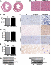
CM-BRCA2−/− mice show no basal cardiac phenotypes. A, representative transverse sections of H&E-stained hearts demonstrate similar morphology at base line between CM-BRCA2−/− mice and their WT littermate controls (n = 4–6/group; age, 10–12 weeks; scale bar, 1 mm). Representative photomicrographs of LV tissues (B) and myocyte cross-sectional area (CSA) (C) were similar between CM-BRCA2−/− mice and their WT littermate controls (n = 4–6/group; 150 cardiomyocytes measured per mouse; age, 10–12 weeks; scale bar, 20 μm). D and E, echocardiographic assessments of left ventricular ejection fraction (LVEF) (D) and fractional shortening (LVFS) (E) show that CM-BRCA2−/− mice and WT littermate controls exhibit similar cardiac function at base line (age, 10–12 weeks; n = 8–9). F, similar extents of cardiac apoptosis (TUNEL and caspase-3 staining (brown)), DNA DSBs (γH2AX focus formation), and RAD51 focusing were seen at base line in CM-BRCA2−/− mice and their WT littermate controls. Spleen (TUNEL) and LV sections (caspase-3, γH2AX, and RAD51) from DOX-treated mice were used as positive (+ve) controls. Blue arrowheads indicate TUNEL-, γH2AX-, and RAD51-positive nuclei (n = 6/group; age, 10–12 weeks; scale bars, 20 μm for TUNEL and caspase-3 micrographs and 10 μm for γH2AX and RAD51). Immunoblots for p53 (G) and ATM (H) from total heart lysates showed similar basal p53 and ATM expressions in the hearts of CM-BRCA2−/− mice and their WT littermate controls. GAPDH was used as a loading control (n = 3–4/group; age, 10–12 weeks). For C, D, and E, data are presented as mean ± S.D.
FIGURE 3.
FIGURE 3.
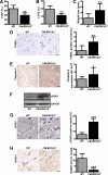
Targeted disruption of BRCA2 in cardiomyocytes exacerbates DOX-induced cardiotoxicity in CM-BRCA2−/− mice. A and B, echocardiographic determination of left ventricular ejection fraction (LVEF) (A) and fractional shortening (LVFS) (B) in CM-BRCA2−/− mice and their WT littermate controls. DOX was administered (10 mg/kg intraperitoneally) 7 days before echocardiographic examination to CM-BRCA2−/− mice and their WT littermate controls (10–12 weeks old). LV ejection fraction and LV fractional shortening values indicate significantly reduced LV performance in CM-BRCA2−/− 7 days post-DOX treatment compared with their WT littermate controls (n = 8–9/group; **, p < 0.01). C, DOX treatment induced significant weight loss in grams (gms) in CM-BRCA2−/− mice in comparison with WT littermate controls 7 days post-DOX treatment (n = 8–9; **, p < 0.001). D and E, cardiomyocyte-specific BRCA2 loss is associated with greater DOX-induced apoptosis, DNA damage (DSBs), and diminished DNA-repair. IHC and quantification of the LV sections from CM-BRCA2−/− mice show increased DOX-induced apoptosis as evident by significantly more TUNEL-positive nuclei (blue arrowhead) (D) and caspase-3 staining (brown) (E) in comparison with the WT littermate controls (n = 6/group; *, p < 0.05; **, p < 0.01; scale bars, 50 μm for caspase-3 and 10 μm for TUNEL). F, immunoblot performed on total heart lysate showed higher γH2AX expression in the samples from CM-BRCA2−/− mice compared with their WT littermate controls. G, representative micrographs and quantification of γH2AX staining of LV sections from DOX-treated CM-BRCA2−/− mice and their WT littermate controls. Significantly higher γH2AX expression (blue arrowheads) was observed in the LV sections of CM-BRCA2−/− mice (n = 6/group; ***, p < 0.001; scale bar, 10 μm). H, IHC staining and quantification for RAD51 foci on LV sections from DOX-treated CM-BRCA2−/− mice and their WT littermate controls showed significantly reduced RAD51 focus formation (blue arrowheads) in the samples from CM-BRCA2−/− mice relative to those from their WT littermate controls (n = 6/group; ***, p < 0.001; scale bar, 10 μm). For A–E and G and H, data are presented as mean ± S.D.
FIGURE 4.
FIGURE 4.
Exacerbated DOX-induced cardiotoxicity in CM-BRCA2−/− mice is p53-mediated. Representative micrograph and quantification of p53 staining (brown staining) (A) and immunoblot for total and phosphorylated (serine 15) p53 (B) showed higher DOX-induced p53 expression and activation in LV sections from DOX-treated CM-BRCA2−/− mice in comparison with those from their WT littermate controls (n = 6/group; **, p < 0.01; scale bar, 50 μm). C and D, immunoblots for PUMA (C) and Bax and Bcl-2 (D) and corresponding quantifications showed increased PUMA expression and elevated Bax/Bcl-2 ratios in the hearts of DOX-treated CM-BRCA2−/− mice in comparison with corresponding values from samples from their WT littermate controls (n = 6/group; *, p < 0.05). E, increased p53, PUMA, and Bax/Bcl-2 ratios were associated with more cytochrome c release in the left ventricle of DOX-treated CM-BRCA2−/− mice as evident by IHC staining (brown) and corresponding quantification (n = 6/group; *, p < 0.05). Immunoblots for total ATM, phosphorylated ATM, and Chk2 (F) as well as p21 (G) and GADD45 (H) on total heart lysates from CM-BRCA2−/− mice and their WT littermate controls show increased expression and activation of ATM (upper band; indicated by arrow), increased activation of Chk2, but similar p21 and GADD45α expression in the hearts of DOX-treated CM-BRCA2−/− mice in comparison with the WT control littermate controls. GAPDH was used as a loading control (n = 3–4/group; age, 10–12 weeks). AU, arbitrary units. For A, D, and E, data are presented as mean ± S.D.

References

    1. Rahman N., Stratton M. R. (1998) The genetics of breast cancer susceptibility. Annu. Rev. Genet. 32, 95–121 - PubMed
    1. Kinzler K. W., Vogelstein B. (1997) Cancer-susceptibility genes. Gatekeepers and caretakers. Nature 386, 761–763 - PubMed
    1. Chen P. L., Chen C. F., Chen Y., Xiao J., Sharp Z. D., Lee W. H. (1998) The BRC repeats in BRCA2 are critical for RAD51 binding and resistance to methyl methanesulfonate treatment. Proc. Natl. Acad. Sci. U.S.A. 95, 5287–5292 - PMC - PubMed
    1. Yoshida K., Miki Y. (2004) Role of BRCA1 and BRCA2 as regulators of DNA repair, transcription, and cell cycle in response to DNA damage. Cancer Sci. 95, 866–871 - PMC - PubMed
    1. Connor F., Bertwistle D., Mee P. J., Ross G. M., Swift S., Grigorieva E., Tybulewicz V. L., Ashworth A. (1997) Tumorigenesis and a DNA repair defect in mice with a truncating Brca2 mutation. Nat. Genet. 17, 423–430 - PubMed

Publication types