. 2012 Jan 27;287(5):3541-9.
doi: 10.1074/jbc.M111.261016. Epub 2011 Dec 8.

The enzymatic activity of lysyl oxidas-like-2 (LOXL2) is not required for LOXL2-induced inhibition of keratinocyte differentiation

Affiliations

The enzymatic activity of lysyl oxidas-like-2 (LOXL2) is not required for LOXL2-induced inhibition of keratinocyte differentiation

Jennie Lugassy et al. J Biol Chem. .

Abstract

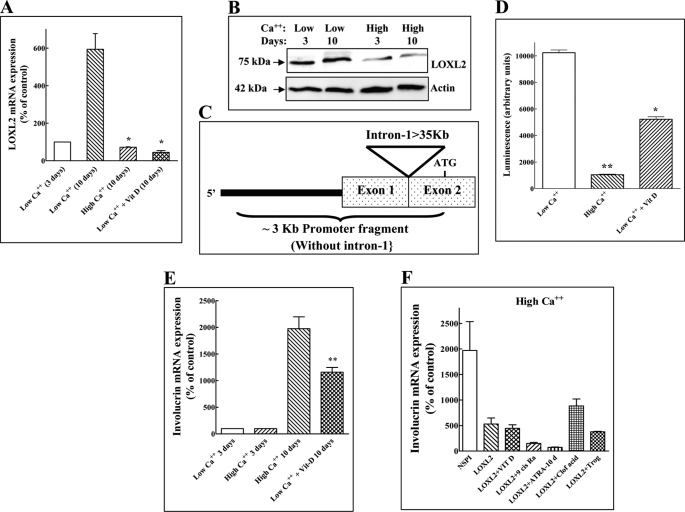
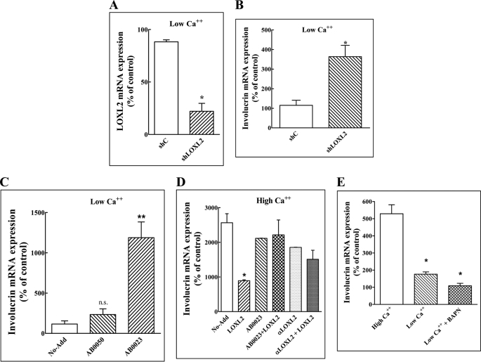
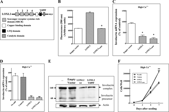
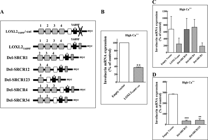
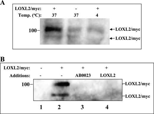
Lysyl oxidase-like-2 (LOXL2) induces tumor progression and fibrosis. It also inhibits the differentiation of keratinocytes promoting development of squamous cell carcinomas. Stimulation of HaCaT skin keratinocytes with exogenous LOXL2 or overexpression of LOXL2 in these cells inhibits their differentiation as manifested by inhibition of calcium or vitamin D-induced involucrin expression. The inhibition was abrogated by the LOXL2 function-blocking monoclonal antibody AB0023 as well as by an anti-LOXL2 polyclonal antibody. Surprisingly, a point-mutated form of LOXL2 (LOXL2(Y689F)) lacking enzymatic activity, as well as a LOXL2 deletion mutant lacking the entire catalytic domain, also inhibited calcium or vitamin D-induced up-regulation of involucrin expression, suggesting that the enzymatic activity of LOXL2 is not required for this activity. This conclusion was supported by experiments that showed that β-aminoproprionitrile, an irreversible competitive inhibitor of the enzymatic activity of all lysyl oxidases, is unable to abolish the LOXL2-induced inhibition of HaCaT cell differentiation. The activity of LOXL2(Y689F) required the presence of the fourth scavenger receptor-cysteine-rich (SRCR) domain of LOXL2, which is also the binding target of AB0023. Epitope-tagged LOXL2(Y689F) was internalized at 37 °C by HaCaT cells. The internalization was inhibited by AB0023 and by competition with unlabeled LOXL2, suggesting that these cells may express a LOXL2 receptor. Our results suggest that agents that inhibit the enzymatic activity of LOXL2 may not suffice to inhibit completely the effects of LOXL2 on complex processes that involve altered states of cellular differentiation.


Figures

FIGURE 1.
FIGURE 1.
LOXL2 inhibits involucrin expression induced by keratinocyte differentiation inducing factors. A, HaCaT cells were cultured in medium containing low or high calcium concentrations. Some low calcium dishes received, in addition, vitamin D (10−8 m) as indicated. After 10 days endogenous LOXL2 mRNA levels were measured using real-time qPCR. The mRNA levels of cells cultured 10 days in the presence of high calcium concentrations or in the presence of vitamin D were compared with the levels of LOXL2 mRNA expression in cells cultured for 10 days in the presence of low calcium concentrations for 10 days. B, cell lysates were prepared from HaCaT cells cultured in low and high Ca2+ after 3 or 10 days as indicated. Aliquots containing equal protein concentrations were subjected to Western blot analysis using polyclonal anti-LOXL2 antibodies. Blots were then stripped and probed for β-actin. C, picture showing the structure of the LOXL2 promoter fragment used. The large intron containing the hypoxia response element (23) downstream to untranslated exon-1 is not included in the promoter fragment. D, the luciferase activity induced by the LOXL2 promoter in HaCaT cells. In response to high calcium concentrations or in response to 10−8 m vitamin D was measured. The y axis represents luciferase activity normalized to a Renilla control. The data shown represent mean values of two independent experiments preformed in triplicate. Means were compared with the low Ca2+ control. E, the effects of calcium and vitamin D on involucrin mRNA expression were measured as described under A for LOXL2 mRNA expression. Means were compared with the high Ca2+ control. F, HaCaT cells cultured in medium containing high calcium concentrations were infected or not with empty lentivirus expression vector (NSPI) or with lentiviruses directing expression of LOXL2Myc (LOXL2) as indicated. Some dishes containing cells infected with lentiviruses directing expression of LOXL2Myc were supplemented with 10−8 m vitamin D (Vit D), 2 μm ATRA (ATRA), 20 μm troglitazone, 2 μm 9-cis-retinoic acid or 400 μm clofibrinic acids. The differentiation inducers were added every other day. Involucrin mRNA expression was measured after 10 days using real-time qPCR and normalized to β-actin mRNA expression.
FIGURE 2.
FIGURE 2.
The lysyl oxidase enzyme activity inhibitor BAPN fails to neutralize the inhibitory effect of LOXL2 on involucrin expression. A, HaCaT cells were cultured in low calcium medium and infected with lentiviruses expressing a non-targeting shRNA (Shc) or with lentiviruses expressing a shRNA directed against LOXL2 (shLOXL2) for 10 days. The LOXL2 mRNA concentrations in the cells were then measured by real-time qPCR. Means were compared with the low Ca2+ (10 days) control. B, the expression of involucrin mRNA was measured in HaCaT cells expressing a non-targeting shRNA (Shc) or a LOXL2 targeting shRNA (shLOXL2) as described under A for the LOXL2 mRNA. C, HaCaT cells were cultured in medium containing low calcium concentrations in the absence of any additions (No-Add), in the presence of 5 μg/ml of a control monoclonal antibody that binds to the enzymatic site of LOXL2 but does not inhibit the enzymatic activity (AB0050) or in the presence of 5 μg/ml of an LOXL2-neutralizing antibody (AB0023), which were replenished every other day. The relative concentrations of the involucrin mRNA were measured using real-time qPCR as described. Means were compared with the control. D, HaCaT cells were cultured in medium containing high calcium concentrations in the absence of any additions (No-Add), 5 μg/ml LOXL2Myc (LOXL2), 20 μg/ml AB0023, or 20 μg/ml of a purified anti-LOXL2 polyclonal antibody (αLOXL2). Some of the wells the received LOXL2 also received antibodies as indicated. Added proteins were replenished every other day. After 10 days the expression of involucrin mRNA was determined by real-time qPCR as described. Means were compared with the control. E, HaCaT cells were cultured in medium containing high or low calcium concentrations as indicated for 10 days. Some of the low calcium wells received in addition BAPN (0.2 mm). Medium and BAPN were replenished every other day. After 10 days involucrin mRNA expression levels were determined using real-time qPCR. Means were compared with the high Ca2+ control.
FIGURE 3.
FIGURE 3.
An enzyme dead LOXL2 point mutant inhibits the differentiation of HaCaT cells. A, a schematic presentation showing the location of the Y689F point mutation introduced into the LOXL2 LTQ domain. B, the enzymatic activity of affinity-purified LOXL2Myc, LOXL2Y689F, or of purified conditioned medium derived from control cells infected with empty lentiviruses and purified identically (Empty vector) were determined using the Amplex Red assay as described under “Experimental Procedures.” The data shown represent mean values of two independent experiments preformed in duplicate. Means were compared with the wild-type LOXL2 Control. C, HaCaT cells were cultured in medium containing high calcium and infected with empty lentiviruses (Empty vector) or lentiviruses directing expression of LOXL2Myc or LOXL2Y689F as indicated. After 10 days involucrin mRNA levels were determined using real-time qPCR. Means were compared with the empty vector control. D, HaCaT cells were grown in medium containing high calcium concentration without additions (No-add), or in medium supplemented with elution buffer (control), purified LOXL2Myc (LOXL2), or LOXL2Y689F (LOXL2Y689F). Added factors were replenished every other day. After 10 days involucrin mRNA levels were determined using real-time qPCR. Means were compared with the control. E, cell lysates were prepared from cells treated as described in D. The presence of involucrin complex and involucrin precursor in the cell lysates was determined by Western blot analysis. F, HaCaT cells were seeded in 24-well plates (5 × 10³ cells/well) in the presence of high calcium concentration. Wells received either no additions (No-add), elution buffer (Vehicle), LOXL2Myc (5 μg/ml), or LOXL2Y689F (5 μg/ml). Added factors were replenished every other day. Adherent cells were counted every other day using a Coulter counter. Means were compared with the low LOXL2 and LOXL2Y689F controls, respectively, on day 7.
FIGURE 4.
FIGURE 4.
The non-enzymatic effects of LOXL2 on the differentiation of HaCaT cells are mediated by the fourth SRCR domain of LOXL2. A, a schematic presentation of the catalytic domain deletion mutant derived from LOXL2Y689F and of the various SRCR deletion mutants of LOXL2Y689F. B–D, HaCaT cells grown in medium containing high calcium concentrations were infected with empty lentiviruses (Empty vector) and with lentiviruses directing expression of LOXL2Y689F/-cat. B, or lentiviruses directing expression of various LOXL2Y689F deletion mutants lacking various SRCR domains as indicated (C and D). The cells were cultured in 6-well plates for 10 days. The data shown represent mean values of two independent experiments preformed in triplicate. The expression of involucrin mRNA was then determined using real-time qPCR. Means were compared with the empty vector control.
FIGURE 5.
FIGURE 5.
Evidence for a putative LOXL2 receptor of HaCaT cells. A, HaCaT cells were cultured in medium containing high calcium concentration to confluence in 10-cm dishes. They were then incubated with or without purified LOXL2Myc as indicated for 0.5 h at 4 °C or at 37 °C. At the end of the incubation the cells were washed and trypsinized to remove cell surface-associated LOXL2Myc. The cells were then lysed, and lysate aliquots containing equal amounts of protein were analyzed by Western blot using an anti-Myc antibody to detect LOXL2Myc. B, HaCaT cells were cultured as described above. Upon reaching confluence the medium was exchanged with conditioned medium (high calcium) derived from HEK293 cells infected with empty lentiviruses (lane 1), with fresh HaCaT growth medium containing high calcium (lanes 2 and 3), or conditioned medium from HEK293 cells producing recombinant untagged LOXL2 (lane 4). LOXL2Myc (2 μg/ml) or AB0023 antibody (20 μg/ml) were added as indicated. After a 30-min incubation at 37 °C the cells were trypsinized and lysed, and LOXL2Myc in cell lysates were visualized as described under A.

References

    1. Saito H., Papaconstantinou J., Sato H., Goldstein S. (1997) Regulation of a novel gene encoding a lysyl oxidase-related protein in cellular adhesion and senescence. J. Biol. Chem. 272, 8157–8160 - PubMed
    1. Lucero H. A., Kagan H. M. (2006) Lysyl oxidase. An oxidative enzyme and effector of cell function. Cell Mol. Life Sci. 63, 2304–2316 - PMC - PubMed
    1. Mäki J. M. (2009) Lysyl oxidases in mammalian development and certain pathological conditions. Histol. Histopathol. 24, 651–660 - PubMed
    1. Tang S. S., Trackman P. C., Kagan H. M. (1983) Reaction of aortic lysyl oxidase with β-aminopropionitrile. J. Biol. Chem. 258, 4331–4338 - PubMed
    1. Rodriguez H. M., Vaysberg M., Mikels A., McCauley S., Velayo A. C., Garcia C., Smith V. (2010) Modulation of lysyl oxidase-like 2 enzymatic activity by an allosteric antibody inhibitor. J. Biol. Chem. 285, 20964–20974 - PMC - PubMed

Publication types

MeSH terms

LinkOut - more resources