. 2012 Feb 15;31(4):870-82.
doi: 10.1038/emboj.2011.449. Epub 2011 Dec 13.

The tudor domain protein kumo is required to assemble the nuage and to generate germline piRNAs in Drosophila

Affiliations

The tudor domain protein kumo is required to assemble the nuage and to generate germline piRNAs in Drosophila

Amit Anand et al. EMBO J. .

Abstract

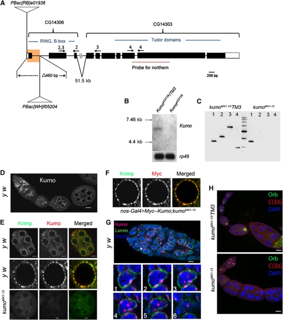
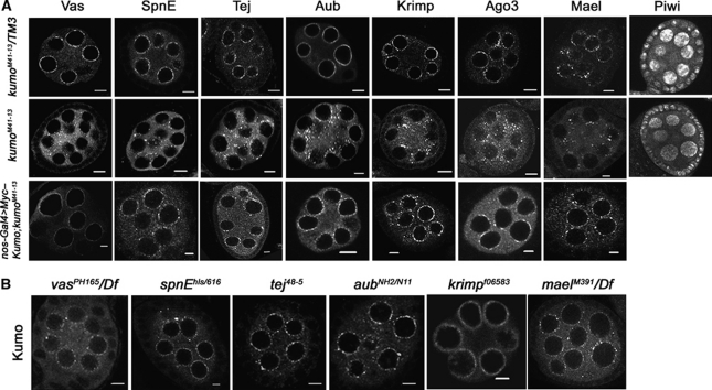
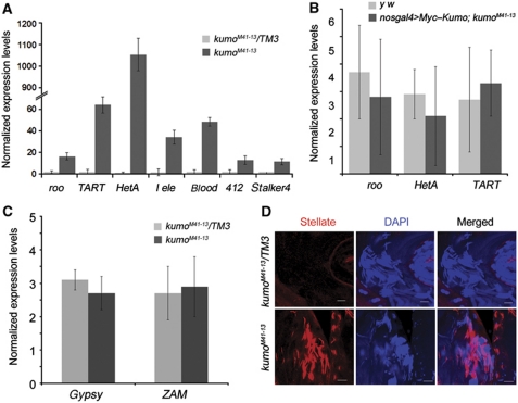
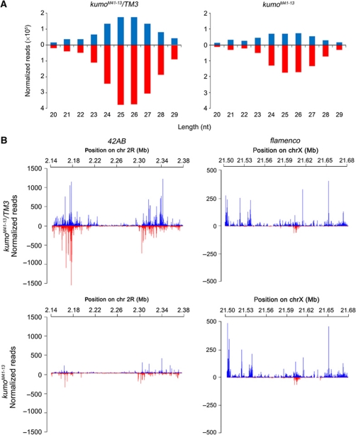
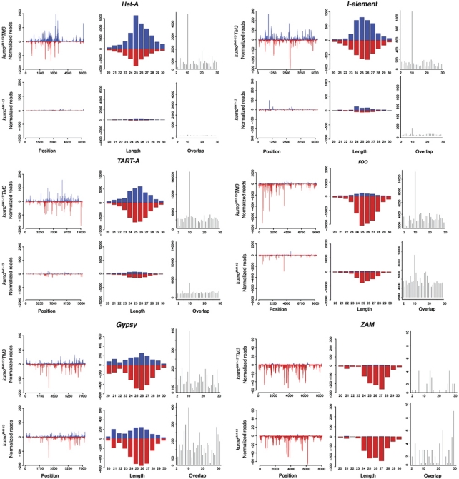
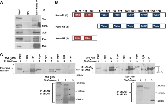
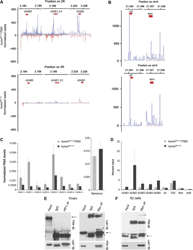
In Drosophila ovaries, distinct Piwi-interacting RNA (piRNA) pathways defend against transposons in somatic and germline cells. Germline piRNAs predominantly arise from bidirectional clusters and are amplified by the ping-pong cycle. In this study, we characterize a novel Drosophila gene, kumo and show that it encodes a conserved germline piRNA pathway component. Kumo contains five tudor domains and localizes to nuage, a unique structure present in animal germline cells, which is considered to be the processing site for germline piRNAs. Transposons targeted by the germline piRNA pathway are derepressed in kumo mutant females. Moreover, germline piRNA production is significantly reduced in mutant ovaries, thereby indicating that kumo is required to generate germline piRNAs. Kumo localizes to the nuage as well as to nucleus early female germ cells, where it is required to maintain cluster transcript levels. Our data suggest that kumo facilitates germline piRNA production by promoting piRNA cluster transcription in the nucleus and piRNA processing at the nuage.


Conflict of interest statement

The authors declare that they have no conflict of interest.

Figures

Figure 1
Figure 1
Kumo localizes to the nuage and nucleus in the germarium and is required for oocyte fate maintenance. (A) Schematic representation of the kumo locus, consisting of two previously annotated genes, CG14303 and CG14306, which are 51.6 kb apart. The region highlighted in yellow represents a deletion of 460 bases in the kumoM41-13 allele by FRT recombination, which encompasses the first exon and the following intron, including the predicted start codon. Positions of PiggyBac insertions are represented in CG14306. The arrows indicate the primer sets used for RT–PCR. (B) Northern blot analysis of the kumo transcript. A transcript of ∼6 kb was detected in the ovarian RNA extracted from the control, but it was not detected in that from kumoe03728 ovaries. (C) RT–PCR with the primer sets denoted in Figure 1A showing the absence of the kumo transcript in kumoM41-13 ovaries. Lane 1, actin control; lanes 2–4, primers against either CG14306 or CG14303 as shown in Figure 1A. (D) y w ovariole immunostained for Kumo showing the expression in germline cells. Perinuclear foci in nurse cells are discernible. Scale bar: 20 μm. (E) (Upper panel) Co-localization of Kumo (red) with a known nuage component, Krimp (green). Scale bar: 5 μm. (Middle panel) Closer view of a single nurse cell nucleus. (Lower panel) Kumo expression is undetectable in a kumoM41-13 egg chamber, and Krimp was mislocalized from the perinuclear nuage. Scale bar: 5 μm. (F) A single nurse cell nucleus showing the co-localization of Myc–Kumo (red) with Krimp (green). (G) (Upper panel) Nuclear localization of Kumo (red, indicated with arrow) in the germarium, which was co-stained for Lamin (green) and DAPI (blue). Scale bar: 10 μm. (Lower panels) Optical sections of germline cells in germarium showing the perinuclear and nuclear foci of Kumo. (H) The control heterozygous and kumoM41-13 ovarioles stained for oocyte markers Orb (green) and C(3)G (red) and DAPI (blue). Orb and C(3)G are undetectable in the egg chambers of stage two and onward in the kumoM41-13 ovary. Scale bar: 10 μm.
Figure 2
Figure 2
kumo genetically interacts with other components of the piRNA pathway. (A) The heterozygous (upper panel) and kumo mutant (middle panel) ovaries immunostained for other nuage components. All of the examined components are mislocalized from the perinuclear nuage in the kumo mutant ovaries. Piwi expression is also slightly reduced in the kumo mutant. Myc–Kumo expression (lower panel) in kumo mutant germline cells rescues the defects in the perinuclear localization of Vas, Tej, Krimp, Aub and Ago3. (B) Immunostaining for Kumo in other nuage component mutant ovaries. Perinuclear localization of Kumo in nurse cells is unaffected in all of the examined mutants. Scale bars: 5 μm.
Figure 3
Figure 3
kumo is involved in the repression of transposons. (A) Quantitative RT–PCR using ovarian RNA extracted from kumo mutant and control ovaries showing the expression levels of representative transposons expressed in the germline. Significant upregulation is seen in the kumo mutant ovaries compared with those in the heterozygous control. (B) Quantitative RT–PCR showing the relative expression levels of roo, HeT-A and TART in ovaries expressing Myc–Kumo in kumo mutant germline cells and control y w ovaries. (C) Quantitative RT–PCR showing no significant differences in the expression levels of gypsy and ZAM transposons, predominantly expressed in somatic cells, between kumoM41-13/TM3 and kumoM41-13 ovaries. The expression levels were normalized with the controls, actin5c and rp49. Error bars indicate the standard deviation of three independent sets of experiments. (D) Immunostaining for Ste (red) with DAPI (blue) showing the upregulation of Ste in kumo mutant testis. Scale bar: 20 μm.
Figure 4
Figure 4
Reduction of germline piRNA levels in kumo mutants. (A) Length histogram of sense and antisense small RNAs produced in the kumo mutant and the control ovaries. An ∼65% reduction in the number of 23–29 nt RNAs was observed in the kumo mutant compared with the control. (B) Diagrams showing piRNA mapping following normalization to a bidirectional cluster at 42AB on chromosome 2R and to unidirectional cluster flamenco on chromosome X. piRNA mapping to the plus strand (blue) and minus strand (red) over cluster 42AB is dramatically reduced in the kumo mutant compared with that in the control ovaries. No such significant reduction in piRNA mapping to the plus strand in the flamenco cluster is observed.
Figure 5
Figure 5
Reduction of piRNA mapping to transposons in kumo mutant ovaries. Mapping of piRNAs to the sense (blue) and antisense (red) strand plotted over the consensus sequence of various transposon families (left panel); length histogram (right) of all matching RNAs from 20 to 30 nt to sense (blue) and antisense (red) strands; and distribution of overlapping piRNAs (right panel) for 1–30 nt. Loss of kumo function depletes piRNA mapping to the sense and antisense strands of Het-A, I-element and TART-A. Concomitantly, piRNA pairs overlapping with 10 nt of those transposons were almost lost in kumo mutant ovaries. A milder reduction in the amount of piRNAs matching to the antisense strand and having a 10-nt overlap was observed for roo. However, for the transposons targeted by somatic piRNAs, a modest reduction in the number of antisense piRNAs mapping to gypsy and no significant difference in piRNAs levels mapping to ZAM were observed in the kumo mutant.
Figure 6
Figure 6
Kumo physically interacts with Vas, SpnE, Aub and Piwi and its interaction with SpnE and Aub is mediated by tudor domains. (A) Western blots showing the co-immunoprecipitation of full-length Myc–Kumo with Vas, Aub, SpnE and Piwi from ovarian lysate. (B) Schematic diagram showing the Kumo variants tagged with FLAG transfected into S2 cells. Kumo-FL, full-length Kumo; Kumo-CT, harbouring all five tudor domains; and Kumo-NT, harbouring RING and B-box domains. (C) Western blots showing the co-immunoprecipitation of FLAG-tagged Kumo variants with Myc–Aub and Myc–SpnE using S2 cell lysate. Both Aub and SpnE are immunoprecipitated with FL- and CT-Kumo but not with NT-Kumo and the control IgG. (Lower panels) Western blots with anti-FLAG showing all examined Kumo variants efficiently pulled down. Asterisks denote nonspecific bands.
Figure 7
Figure 7
The kumo mutation results in reduced cluster transcription and high HP1 occupancy at piRNA clusters. (A, B) Mapping of piRNAs to the indicated regions of the bidirectional cluster at 42AB and the unidirectional clusters at X (cluster 2 and flamenco), which were examined for the expression of the cluster transcripts and HP1 binding. In the kumo mutant ovaries, piRNA mapping to the bidirectional cluster were nearly eliminated, whereas no significant impact was observed in the number of piRNAs matching cluster 2 and flamenco. (C) Strand-specific quantitative RT–PCR showing the expression levels of cluster transcripts from the plus and minus strands (indicated by + and −, respectively) from the 42AB piRNA cluster. RNA levels from both strands in the kumo mutant ovaries are reduced compared with those in the heterozygous control. However, no significant difference in the expression levels of transcripts from a region of the flamenco piRNA cluster was observed between the control and kumo mutant ovaries. Error bars indicate standard error for three independent experiments. (D) Quantification of chromatin immunoprecipitation with anti-HP1 using primers at various regions of the 42AB piRNA cluster in control and kumo mutant ovaries. The percent input of immunoprecipitates is shown for each primer set. HP1 binding was enriched in kumo mutant ovaries compared with that in the kumo heterozygous controls with 1.5-, 12.8-, 3.1-, 2.8- and 4.8-fold increases at 42AB1–5, respectively. No significant increase was observed at unidirectional piRNA clusters, cluster 2 and flamenco, the controls, euchromatin region, rp49 and heterochromatin region (X-TAS) in the kumo mutant. Error bars indicate standard error from two independent experiments. (E) Co-immunoprecipitation of Myc–Kumo and HP1 from the ovarian lysates. Western blots for Myc–Kumo and HP1: 5% of input was loaded on the blots to detect HP1 in Myc–Kumo immunoprecipitate and in the reciprocal immunoprecipitation 1% of input was used. (F) HP1 is co-immunoprecipitated with Myc–Kumo in S2 cells. 1% input was used in the western blot. Asterisks denote the non-specific bands.

Comment in

References

    1. Aravin AA, Klenov MS, Vagin VV, Bantignies F, Cavalli G, Gvozdev VA (2004) Dissection of a natural RNA silencing process in the Drosophila melanogaster germ line. Mol Cell Biol 24: 6742–6750 - PMC - PubMed
    1. Aravin AA, Naumova NM, Tulin AV, Vagin VV, Rozovsky YM, Gvozdev VA (2001) Double-stranded RNA-mediated silencing of genomic tandem repeats and transposable elements in the D. melanogaster germline. Curr Biol 11: 1017–1027 - PubMed
    1. Brennecke J, Aravin AA, Stark A, Dus M, Kellis M, Sachidanandam R, Hannon GJ (2007) Discrete small RNA-generating loci as master regulators of transposon activity in Drosophila. Cell 128: 1089–1103 - PubMed
    1. Chen Y, Pane A, Schupbach T (2007) Cutoff and aubergine mutations result in retrotransposon upregulation and checkpoint activation in Drosophila. Curr Biol 17: 637–642 - PMC - PubMed
    1. Eddy EM (1974) Fine structural observations on the form and distribution of nuage in germ cells of the rat. Anat Rec 178: 731–757 - PubMed

Publication types