. 2012 Apr;25(4):602-14.
doi: 10.1038/modpathol.2011.185. Epub 2011 Dec 9.

Embryonal rhabdomyosarcoma of the uterine cervix: a report of 14 cases and a discussion of its unusual clinicopathological associations

Affiliations

Embryonal rhabdomyosarcoma of the uterine cervix: a report of 14 cases and a discussion of its unusual clinicopathological associations

Louis P Dehner et al. Mod Pathol. 2012 Apr.

Abstract

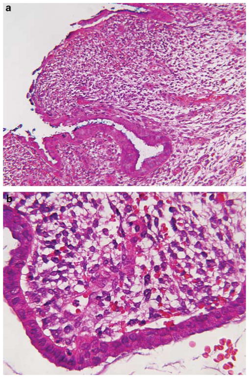
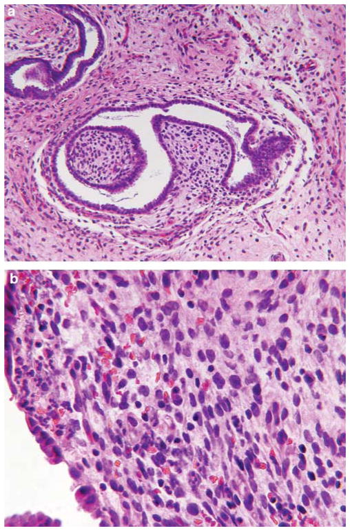
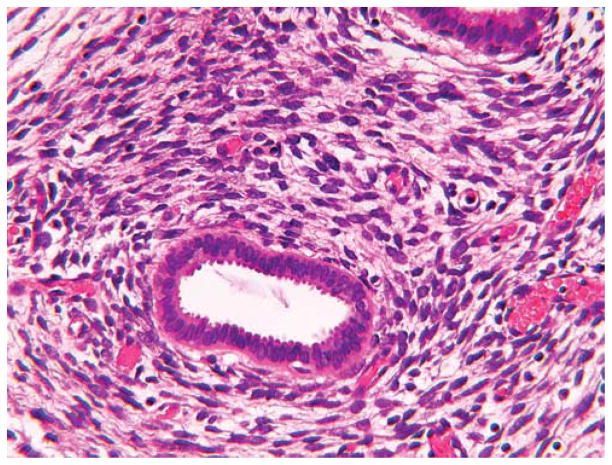
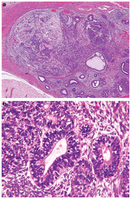
Embryonal rhabdomyosarcoma of the uterine cervix is an uncommon presentation of the most common soft-tissue sarcoma in the first decades of life. Unlike embryonal rhabdomyosarcoma in other anatomic sites, in which 70-80% of cases present before 9 years of age, the average age in our series of 14 cervical cases was 12.4 years (median, 13 years), with an age range of 9 months to 32 years at diagnosis. Of the 14 cases, 12 presented as a polyp at the cervical os; two patients had an infiltrative mass in the cervix without a botryoid polyp. The polyps measured 1.5-5 cm and all had the histopathological pattern of the sarcoma botryoides variant of embryonal rhabdomyosarcoma, with condensations of primitive and differentiated rhabdomyoblasts beneath the surface epithelium and around endocervical glands. Nodules of benign-appearing cartilage were present in the stroma of six cases (43%). One of the embyronal rhabdomyosarcomas from the youngest patient, 9 months old, also had a distinctive microscopic focus of immature tubular profiles in a primitive stroma; these tubules expressed epithelial and neuroendocrine markers. Two patients had a pleuropulmonary blastoma, one diagnosed 9 years before the embryonal rhabdomyosarcoma of the cervix and the other recognized synchronously. This latter 9-year old had a DICER1 germline mutation. One patient presented with hirsutism and had a Sertoli-Leydig cell tumor, an incidentally detected cervical embryonal rhabdomyosarcoma, and nodular hyperplasia of the thyroid. Although a pleuropulmonary blastoma was not documented in the latter patient, ovarian sex-cord stromal tumors and nodular hyperplasia of the thyroid are manifestations of the pleuropulmonary blastoma family tumor and dysplasia syndrome (OMIM 601200). Embryonal rhabdomyosarcoma of the cervix must be distinguished from other rare entities, including adenosarcoma, malignant mixed Mullerian tumor and low-grade stromal sarcoma, as the former has a better prognosis; 12 of our 14 patients remain disease-free following conservative surgery and chemotherapy. Our study suggests that cervical embryonal rhabdomyosarcoma may be another pathological manifestation in the spectrum of extrapulmonary pathology in the setting of pleuropulmonary blastoma.


Conflict of interest statement

Disclosure/conflict of interest The authors declare no conflict of interest.

Figures

Figure 1
Figure 1
(a) Embryonal rhabdomyosarcoma of the cervix in a 13-year-old female patient (Case 9) shows erosion of the surface epithelium and interstitial hemorrhage, which can obscure the underlying pathology. (b) Contiguous areas of the same polyp with intact surface epithelium demonstrated to better advantage the primitive rhabdomyoblasts.
Figure 2
Figure 2
(a) This 9-year-old female (Case 11) presented with a ‘vaginal polyp’, which was traced into the endocervix. Adjacent endocervical glands are surrounded in part by subepithelial tumor cells. Note that the stroma beyond the glands has a hypocellular myxoid appearance. (b) The rhabdomyoblastic nature of the tumor cells is noted by the small-rounded to spindle-shaped tumor cells with delicate tails of eosinophilic cytoplasm.
Figure 3
Figure 3
The nodules of cartilage varied in size and degree of cellularity. (a) This nodule of cartilage was one of several within the stroma (Case 11). The adjacent endocervical gland provides some perspective on its size. (b) A smaller, more cellular nodule is adjacent to an endocervical gland with accompanying embryonal rhabdomyosarcoma.
Figure 4
Figure 4
The dense circumferential pattern of rhabdomyoblasts around endocervical glands was a consistent feature in these polypoid neoplasms (Case 9), regardless of age at diagnosis.
Figure 5
Figure 5
(a) A distinct focus of tumor was present in a sarcoma botryoides of the cervix in a 9-month-old female patient (Case 9). The embryonal rhabdomyosarcomatous pattern is depicted in part in Figure 4. (b) The primitive tubular profiles are surrounded by smaller rosette-like structures in a background of immature, non-rhabdomyosarcomatous mesenchyme.
Figure 6
Figure 6
One case presented as a cervical mass rather than a polyp in a 15-year-old female patient (Case 8). This neoplasm displayed a variety of patterns. (a) An endocervical gland is surrounded in part by embryonal rhabdomyoblasts. (b) Other foci were represented by spindle cell rhabdomyosarcoma. (c) Yet another pattern of this neoplasm had a more blastematous appearance. (d) Anaplastic rhabdomyoblasts were found in several foci. The composite patterns in this neoplasm have a similarity to the histological collage, which is seen in the solid areas of the pleuropulmonary blastoma.
Figure 7
Figure 7
Embryonal rhabdomyosarcoma presented as a hemorrhagic, necrotic polyp from the cervix of this 32-year-old female patient (Case 14). (a) This tumor displayed a range of features from primitive rhabdomyoblasts to well-differentiated rhabdomyoblasts with myotube formation. (b) Pleomorphic tumor cells accompanied rhabdomyoblasts in areas of infiltration into the smooth muscle of cervix. (c) Desmin expression was consistently expressed in the primitive rhabdomyoblasts and differentiated myotubes, but less consistently in the large pleomorphic tumor cells. (d) Nuclear positivity for myogenin was seen in rhabdomyoblasts, but infrequently in the pleomorphic cells.
Figure 8
Figure 8
(a) A large multicystic lesion in the right upper lobe was discovered during the non-invasive staging in this 9-year-old female patient (Case 11). (b) The resected lobe contained a 7.5 × 6.5 × 6 cm3 multicystic lesion, which is seen in the unopened specimen before its collapse after the release of clear fluid. The septa had a smooth translucent appearance and no areas of gross thickening or masses were identified.
Figure 9
Figure 9
(a) Cystic pleuropulmonary blastoma (type I) was composed of cysts, some with a delicate thread-like quality and others with a more prominent fibrous stroma. (b) The cysts were lined by a low cuboidal epithelium and a pale hypocellular stroma. Condensations of primitive round cells or rhabdomyoblasts were not identified after extensive sampling.
Figure 10
Figure 10
(a) Sertoli–Leydig cell tumor of the right ovary shows the presence of discrete solid nests in a hemorrhagic background. (b) Elsewhere, cords of Leydig cells are present in the fibromyxoid stroma.
Figure 11
Figure 11
This tumor occurrence in the oldest patient in this series, a 32-year-old woman (Case 14). (a) A mantle of desmin-positive rhabdomyoblasts was surrounding an endocervical gland. (b) Nuclear staining for myogenin was present in most, but not all, rhabdomyoblasts, which is relatively common in embryonal rhabdomyosarcoma.
Figure 12
Figure 12
This polypoid lesion presented in the cervix in a 9-month-old female (Case 9), the youngest patient in this series. (a) The discrete island of tubules and mesenchyme in this neoplasm contains tubular profiles, which are pancytokeratin positive. (b) Many of these same tubular structures are neuron-specific enolase (shown) and synaptophysin positive. (c) All of the primitive small tumor cells are positive for vimentin and a fraction are positive for desmin (shown). (d) Nuclear positivity for myogenin is seen in a population of primitive small cells without obvious rhabdomyoblastic differentiation.

Similar articles

Cited by

References

    1. Ognjanovic S, Linabery AM, Charbonneau B, et al. Trends in childhood rhabdomyosarcoma incidence and survival in the United States, 1975–2005. Cancer. 2009;115:4218–4226. - PMC - PubMed
    1. Dehner LP. Soft tissues. In: Stocker TJ, Dehner LP, Husain A, editors. Stocker and Dehner’s Pediatric Pathology. 3. Wolter Kluwer–Lippincott Williams & Wilkins; Philadelphia: 2011. pp. 1079–1087.
    1. Leuschner I, Harms D, Mattke A, et al. Rhabdomyosarcoma of the urinary bladder and vagina. A clinicopathologic study with emphasis on recurrent disease: a report from the Kiel Pediatric Tumor Registry and the German CWS Study. Am J Surg Pathol. 2001;25:856–864. - PubMed
    1. Walterhouse DO, Meza JL, Breneman JC, et al. Local control and outcome in children with localized vaginal rhabdomyosarcoma: a report from the Soft Tissue Sarcoma Committee of the Children’s Oncology Group. Pediatr Blood Cancer. 2011;57:76–83. - PMC - PubMed
    1. Herzog CE. Overview of sarcomas in the adolescent and young adult population. J Pediatr Hematol Oncol. 2005;27:215–218. - PubMed

MeSH terms

Supplementary concepts