. 2011 Dec 8;7(1):1-12.
doi: 10.1038/nprot.2011.421.

SILEC: a protocol for generating and using isotopically labeled coenzyme A mass spectrometry standards

Affiliations

SILEC: a protocol for generating and using isotopically labeled coenzyme A mass spectrometry standards

Sankha S Basu et al. Nat Protoc. .

Abstract

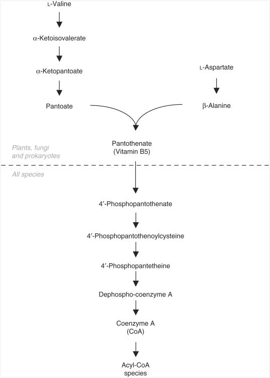
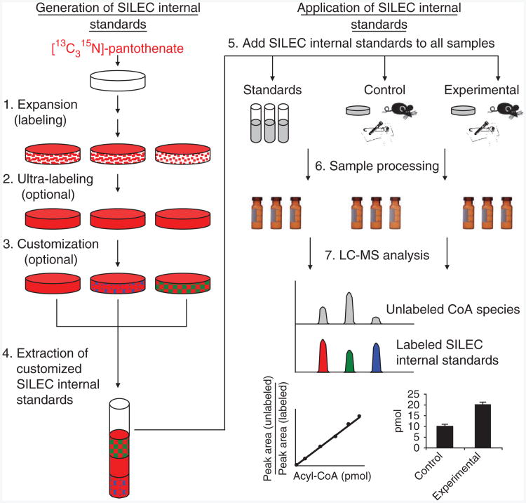
Stable isotope labeling by essential nutrients in cell culture (SILEC) was recently developed to generate isotopically labeled coenzyme A (CoA) and short-chain acyl-CoA thioesters. This was accomplished by modifying the widely used technique of stable isotope labeling by amino acids in cell culture to include [(13)C(3)(15)N]-pantothenate (vitamin B(5)), a CoA precursor, instead of the isotopically labeled amino acids. The lack of a de novo pantothenate synthesis pathway allowed for efficient and near-complete labeling of the measured CoA species. This protocol provides a step-by-step approach for generating stable isotope-labeled short-chain acyl-CoA internal standards in mammalian and insect cells as well as instructions on how to use them in stable isotope dilution mass spectrometric-based analyses. Troubleshooting guidelines, as well as a list of unlabeled and labeled CoA species, are also included. This protocol represents a prototype for generating stable isotope internal standards from labeled essential nutrients such as pantothenate. The generation and use of SILEC standards takes approximately 2-3 weeks.


Figures

Figure 1
Figure 1
LC-MS analysis of acyl-CoA thioesters. (a) Generalized CoA structure, showing the CID-induced neutral loss of the ATP moiety (m/z = 507). The substituent (R group) can include any thioester or derivative of CoA arising from endogenous or exogenous sources (see supplementary table 1 for a list of typical endogenous CoA species). (b) LC-constant neutral loss/MS scans (m/z = 507) of short-chain acyl-CoA species extracted from unlabeled Hepa 1c1c7 mouse hepatoma cells. The extraction was performed as described in the accompanying protocol using the specific LC-MS parameters provided in Figure 2. The precursor parent molecules of various short-chain acyl-CoA species are annotated in the spectrum. c.p.s., counts per second.
Figure 2
Figure 2
LC-MS parameters used in our laboratory for the analysis of short-chain acyl-CoA thioesters.
Figure 3
Figure 3
Pantothenate and coenzyme A biosynthesis. Plants, fungi and many prokaryotes are capable of de novo pantothenate synthesis from valine and aspartate, whereas animals require it as part of their diet.
Figure 4
Figure 4
General scheme for stable isotope labeling by essential nutrients in cell culture (SILEC). (1) Expansion: cells are serially passaged and expanded in the presence of labeled pantothenate. (2) Ultra-labeling: medium containing less serum and more labeled pantothenate is used to increase purity of labeled CoA. (3) Customization: labeled cells are subjected to different biological, pharmaceutical or toxicological exposures to modify the CoA derivative profile within the cells. (4) Extraction: cells are lysed and pooled together to generate a SILEC CoA internal standard mix with a more global CoA profile. (5) SILEC spike-in: equal amounts of SILEC internal standards are spiked into CoA standards, as well as in vitro, in vivo or into clinical experimental samples. (6) Sample processing: standards and samples are subjected to the same sample extraction procedures. (7) LC-MS analysis: CoA species are separated and analyzed by LC-SRM/MS. Co-eluting SILEC internal standards are used to confirm the identity and normalize for the different CoA species.
Figure 5
Figure 5
SILEC labeling and ‘customization’ of CoA metabolome. (ac) LC–neutral loss/MS scans (m/z = 507) of acid-extracted acyl-CoA species in SILEC-labeled Hepa 1c1c7 cells that were untreated (a), treated with 10 mM propionate for 1 h (b) or treated with 10 mM β-hydroxybutyrate for 1 h (c). (df) LC–neutral loss/MS scans (m/z = 507) of acid-extracted acyl-CoA species in Drosophila S2 cells that were untreated (d), treated with 10 mM propionate for 1 h (e) or treated with 10 mM β-hydroxybutyrate for 1 h (f). The precursor ion masses of various labeled short-chain acyl-CoA species can be identified: [13C315N]-acetyl-CoA (m/z = 814), [13C315N]-succinyl-CoA (m/z = 872), [13C315N]-CoASH (m/z = 772), [13C315N]-propionyl-CoA (m/z = 828) and [13C315N]-β-hydroxybutyryl-CoA (m/z = 858). These ‘customized’ CoA extracts can be pooled together to generate a more comprehensive CoA profile. Extraction was performed as specified in the protocol (Fig. 2).
Figure 6
Figure 6
Biosynthetic generation of isotopically labeled menadione-CoA. (a) LC–neutral loss/MS scan (m/z = 507) of acid-extracted CoA species from Hepa 1c1c7 cells treated with 20 μM menadione for 1 h. (b) LC-SRM/MS chromatogram of [13C315N]-menadione-CoA (m/z = 942 → 435) derived from the same extract. Extraction and analysis were performed as specified. By spiking this extract into experimental samples, this isotopically labeled analyte can be used as an internal standard for quantifying unlabeled menadione-CoA.
Figure 7
Figure 7
LC-SRM/MS chromatograms of CoASH harvested from [13C315N]-pantothenate-labeled cells. Both unlabeled CoASH (m/z = 768 → 261) and [13C315N]-CoASH (m/z = 772 → 265) chromatograms are presented to illustrate the labeling efficiency. (a) Suboptimal labeling: the amount of unlabeled CoASH was 5.7%. (b) Near-complete labeling: the amount of unlabeled CoASH was 0.5%. Extraction and analysis were performed as specified in the protocol.

References

    1. Hsieh Y. HPLC-MS/MS in drug metabolism and pharmacokinetic screening. Expert Opin Drug Metab Toxicol. 2008;4:93–101. - PubMed
    1. Brown SC, Kruppa G, Dasseux JL. Metabolomics applications of FT-ICR mass spectrometry. Mass Spectrom Rev. 2005;24:223–231. - PubMed
    1. Hu QZ, et al. The Orbitrap: a new mass spectrometer. J Mass Spectrom. 2005;40:430–443. - PubMed
    1. Whalen K, Gobey J, Janiszewski J. A centralized approach to tandem mass spectrometry method development for high-throughput ADME screening. Rapid Commun Mass Spectrom. 2006;20:1497–1503. - PubMed
    1. Taylor CF, et al. A systematic approach to modeling, capturing, and disseminating proteomics experimental data. Nat Biotechnol. 2003;21:247–254. - PubMed

Publication types