

Osteonecrosis in children after allogeneic hematopoietic cell transplantation: study of prevalence, risk factors and longitudinal changes using MR imaging
- PMID: 22158389
- PMCID: PMC3310343
- DOI: 10.1038/bmt.2011.234
Osteonecrosis in children after allogeneic hematopoietic cell transplantation: study of prevalence, risk factors and longitudinal changes using MR imaging
Abstract
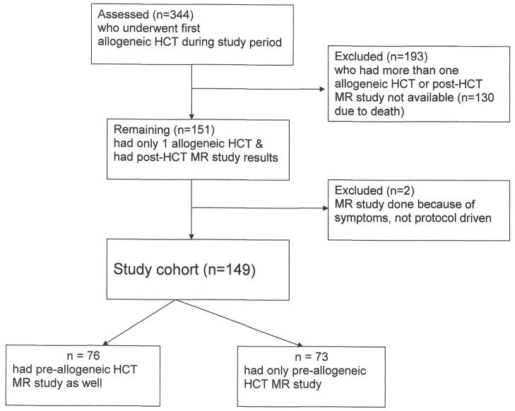
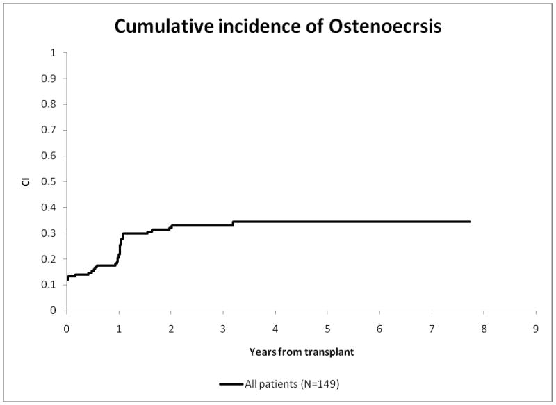
Osteonecrosis after hematopoietic SCT (HCT) has seldom been addressed in pediatric populations. At our institution, since January 2002, children undergoing allogeneic HCT (alloHCT) receive yearly follow-up magnetic resonance imaging (MR) of hips and knees. To estimate the prevalence, longitudinal changes and associated risk factors for osteonecrosis after alloHCT, we reviewed MRs for children who underwent single alloHCT during the study period. We analyzed 149 of 344 patients who had post-HCT MR imaging performed (84 males; median age 11 years (range, 0.5-21 years)), median follow-up time was 32.6 months (range, 2.8-97.2 months). In all, 44 (29.5%) developed osteonecrosis of hips and/or knees; of those, 20 (45%) had at least 30% epiphyseal involvement. In 23 (52%), osteonecrosis lesions were identified in the first and in 43 (98%) by the third yearly scan. Knees were more frequently involved than hips; severity of osteonecrosis was greater in hips. Those who had pre-alloHCT osteonecrosis, two patients' hips and six patients' knees resolved completely; three patients' osteonecrosis lesions regressed after alloHCT. On risk factor analysis, age at time of alloHCT (P=0.051) and osteonecrosis identified by MRs before alloHCT (P=0.001) were the primary risk factors. This analysis shows that preventive strategies for osteonecrosis in this population should focus on measures to minimize risk factors before alloHCT.
Figures
References
-
- Socie G, Cahn JY, Carmelo J, et al. Avascular necrosis of bone after allogeneic bone marrow transplantation: analysis of risk factors for 4388 patients by the Societe Francaise de Greffe de Moelle (SFGM) Br J Haematol. 1997;97(4):865–870. - PubMed
-
- Enright H, Haake R, Weisdorf D. Avascular necrosis of bone: a common serious complication of allogeneic bone marrow transplantation. Am J Med. 1990;89(6):733–738. - PubMed
-
- Karimova EJ, Rai SN, Howard SC, et al. Femoral head osteonecrosis in pediatric and young adult patients with leukemia or lymphoma. J Clin Oncol. 2007;25(12):1525–1531. - PubMed
Publication types
MeSH terms
Grants and funding
LinkOut - more resources
Full Text Sources
Medical
