. 2012 Jan 27;287(5):3138-46.
doi: 10.1074/jbc.M111.277921. Epub 2011 Dec 9.

Apoptosis-dependent externalization and involvement in apoptotic cell clearance of DmCaBP1, an endoplasmic reticulum protein of Drosophila

Affiliations

Apoptosis-dependent externalization and involvement in apoptotic cell clearance of DmCaBP1, an endoplasmic reticulum protein of Drosophila

Ryo Okada et al. J Biol Chem. .

Abstract

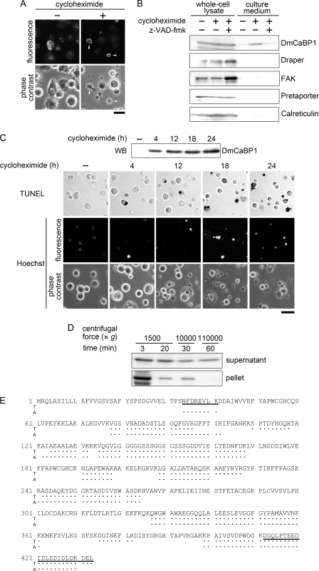
To elucidate the actions of Draper, a receptor responsible for the phagocytic clearance of apoptotic cells in Drosophila, we isolated proteins that bind to the extracellular region of Draper using affinity chromatography. One of those proteins has been identified to be an uncharacterized protein called Drosophila melanogaster calcium-binding protein 1 (DmCaBP1). This protein containing the thioredoxin-like domain resided in the endoplasmic reticulum and seemed to be expressed ubiquitously throughout the development of Drosophila. DmCaBP1 was externalized without truncation after the induction of apoptosis somewhat prior to chromatin condensation and DNA cleavage in a manner dependent on the activity of caspases. A recombinant DmCaBP1 protein bound to both apoptotic cells and a hemocyte-derived cell line expressing Draper. Forced expression of DmCaBP1 at the cell surface made non-apoptotic cells susceptible to phagocytosis. Flies deficient in DmCaBP1 expression developed normally and showed Draper-mediated pruning of larval axons, but a defect in the phagocytosis of apoptotic cells in embryos was observed. Loss of Pretaporter, a previously identified ligand for Draper, did not cause a further decrease in the level of phagocytosis in DmCaBP1-lacking embryos. These results collectively suggest that the endoplasmic reticulum protein DmCaBP1 is externalized upon the induction of apoptosis and serves as a tethering molecule to connect apoptotic cells and phagocytes for effective phagocytosis to occur.


Figures

FIGURE 1.
FIGURE 1.
Identification and expression pattern of DmCaBP1. A, whole-cell lysates of S2 cells were subjected to affinity chromatography using Sepharose conjugated with the extracellular region of Draper fused to GST (Draper-GST) or GST alone. The bound materials were eluted and analyzed by SDS-PAGE followed by staining with silver. The arrowhead points to the position of a band that was subsequently identified to be DmCaBP1. B, the entire amino acid sequence of DmCaBP1 deduced from the nucleotide sequence of cDNA is shown with the positions of peptides identified in the MS analysis (underlined) as well as predicted domain structures. The numbering denotes amino acid positions with the N terminus as 1. The boxed, colored (blue and red), and bolded/shaded regions indicate the signal peptide, the thioredoxin-like domains, and the ER-retention motif, respectively. Refer to supplemental Fig. S1 for more information. C, the binding of DmCaBP1 to Draper was examined by an ELISA-like solid-phase assay. GST-fused Draper or GST alone was incubated in a culture container that had been coated with the indicated MBP-fused proteins, and the amount of GST proteins remaining in the container after washing was determined by an immunochemical reaction. The mean ± S.D. of the data (n = 5) from one of two independent experiments with similar results are presented. D, lysates of whole animals at the indicated developmental stages were analyzed by Western blotting for the level of DmCaBP1. E, dispersed embryonic cells (stage 16) of a fly line that expresses YFP fused to the ER-retention motif KDEL at the C terminus as a marker for ER-residing proteins (ER marker) were immunocytochemically analyzed using anti-DmCaBP1 antibody for the subcellular localization of DmCaBP1. Phase-contrast and fluorescence views of the same microscopic fields are shown as laterally aligned panels. The inset shows a magnified view of a cell pointed by an arrowhead in each panel. Scale bar = 20 μm.
FIGURE 2.
FIGURE 2.
Apoptosis-dependent externalization of DmCaBP1. A, S2 cells before and after the treatment with cycloheximide were immunocytochemically analyzed for the subcellular localization of DmCaBP1. Fluorescence and phase-contrast views of the same microscopic fields are shown as vertically aligned panels. The arrowheads indicate the cells where the localization of DmCaBP1 has been altered. Scale bar = 10 μm. B, S2 cells were treated with cycloheximide in the absence and presence of the caspase inhibitor z-VAD-fmk, and their whole-cell lysates as well as the culture media were analyzed by Western blotting for the levels of the indicated proteins. Only portions of the gel containing the signals derived from the corresponding proteins are shown. FAK, focal adhesion kinase. C, S2 cells were maintained in the presence of cycloheximide for the indicated periods of time. The culture media were analyzed for the level of DmCaBP1 by Western blotting (WB), whereas the cells were subjected to cytochemical analyses for the occurrence of DNA cleavage (TUNEL) and chromatin condensation (Hoechst). In the analysis of DNA cleavage, TUNEL-positive nuclei are shown in black. In the analysis of chromatin condensation, fluorescence and phase-contrast views of the same microscopic field are shown as vertically aligned panels. Scale bar = 20 μm. D, culture media of cycloheximide-treated S2 cells were first centrifuged at 1,500 × g for 3 min, and the resulting supernatants were recentrifuged at 1,500 × g for 20 min. Supernatants were collected and centrifuged at 10,000 × g for 30 min, and the resulting supernatants were recentrifuged at 110,000 × g for 60 min. Supernatants and pellets obtained after each centrifugation were analyzed by Western blotting for the presence of DmCaBP1. E, DmCaBP1 present in the culture media of cycloheximide-treated S2 cells and whole-cell lysates of normal S2 cells was immunoprecipitated with anti-DmCaBP1 antibody and subjected to the MS analysis after digestion with trypsin (T) or Achromobacter protease I (A). Peptides identified in both DmCaBP1 preparations are shown with dots along with the amino acid sequence of DmCaBP1 deduced from the nucleotide sequence of its cDNA. Peptides including the presumed N terminus and C terminus of mature DmCaBP1 are underlined. Refer to supplemental Fig. S3 for more information.
FIGURE 3.
FIGURE 3.
Surface DmCaBP1-mediated phagocytosis of latex beads and non-apoptotic cells. A, cycloheximide-treated S2 cells (left panel) and l(2)mbn cells (right panel) were incubated with recombinant DmCaBP1 proteins fused to GST and MBP, respectively. GST alone and MBP-βgal were included as controls for GST-DmCaBP1 and MBP-DmCaBP1-βgal, respectively. The cells were recovered by centrifugation, and their whole-cell lysates were examined by Western blotting using anti-GST antibody (left panel) or anti-MBP antibody (right panel) together with the input proteins, GST-DmCaBP1/GST (left panel) and MBP-DmCaBP1-βgal/MBP-βgal (right panel). B, latex beads coated with GST-fused DmCaBP1 (GST-DmCaBP1) or GST alone were subjected to an assay for phagocytosis with l(2)mbn cells that had been treated with double-stranded RNA (dsRNA) containing mRNA sequences of the indicated genes, as phagocytes. The level of phagocytosis is shown relative to that of non-coated latex beads, taken as 100. NS, not significant. The mean ± S.D. of the data (n = 6) from one of three independent experiments with similar results are presented. C, S2 cells transfected with DNA for the expression of GPI-anchored DmCaBP1 (GPI-DmCaBP1) or with the vector alone were subjected to an immunofluorescence analysis with anti-DmCaBP1 antibody under a membrane-non-permeabilized condition. Scale bar = 10 μm. D, S2 cells analyzed in B as well as untransfected cells (none) were examined for susceptibility to phagocytosis by l(2)mbn cells. Data are expressed as the mean ± S.D. of the results from three independent experiments (n = 3 in each experiment).
FIGURE 4.
FIGURE 4.
Involvement of DmCaBP1 in apoptotic cell clearance in Drosophila. A, whole-animal lysates of adult flies generated by the mobilization of P-element were analyzed by Western blotting for the level of DmCaBP1. DmCaBP1H20 and DmCaBP1Δ1 are fly lines obtained through the precise and imprecise excision of P-element in the original fly line, respectively. Refer to supplemental Fig. S4 for more information. B, dispersed cells prepared from stage-16 embryos of DmCaBP1H20 and DmCaBP1Δ1 were examined for the level of phagocytosis. Data are expressed as the mean ± S.D. of the results from three independent experiments (n = 5 in each experiment). C, brains dissected from DmCaBP1H20 and DmCaBP1Δ1 as well as Draper-lacking (drprΔ5) flies at the indicated developmental stages (APF, after pupation formation) were histochemically examined for the existence of γ neuron axons. The arrowheads point to the axons removed during metamorphosis. Scale bar = 20 μm.
FIGURE 5.
FIGURE 5.
Functional relationship between DmCaBP1 and Pretaporter. A, lysates of adult flies of the indicated lines were analyzed by Western blotting for the levels of DmCaBP1, Pretaporter, and Draper. The fly line prtpΔ1 is a null-mutant for pretaporter (17). B, dispersed cells prepared from stage-16 embryos of the indicated fly lines were examined for the level of phagocytosis. Data are expressed as the mean ± S.D. of the results from three independent experiments (n = 5 in each experiment). NS, not significant.

Similar articles

Cited by

References

    1. Liao D. J. (2005) The scavenger cell hypothesis of apoptosis. Apoptosis redefined as a process by which a cell in living tissue is destroyed by phagocytosis. Med. Hypotheses 65, 23–28 - PubMed
    1. Nakanishi Y., Nagaosa K., Shiratsuchi A. (2011) Phagocytic removal of cells that have become unwanted. Implications for animal development and tissue homeostasis. Dev. Growth Differ. 53, 149–160 - PubMed
    1. Savill J., Fadok V. (2000) Corpse clearance defines the meaning of cell death. Nature 407, 784–788 - PubMed
    1. Lauber K., Blumenthal S. G., Waibel M., Wesselborg S. (2004) Clearance of apoptotic cells. Getting rid of the corpses. Mol. Cell 14, 277–287 - PubMed
    1. Ravichandran K. S., Lorenz U. (2007) Engulfment of apoptotic cells. Signals for a good meal. Nat. Rev. Immunol. 7, 964–974 - PubMed

Publication types

MeSH terms