Identification of functionally distinct TRAF proinflammatory and phosphatidylinositol 3-kinase/mitogen-activated protein kinase/extracellular signal-regulated kinase kinase (PI3K/MEK) transforming activities emanating from RET/PTC fusion oncoprotein
- PMID: 22158616
- PMCID: PMC3281745
- DOI: 10.1074/jbc.M111.322677
Identification of functionally distinct TRAF proinflammatory and phosphatidylinositol 3-kinase/mitogen-activated protein kinase/extracellular signal-regulated kinase kinase (PI3K/MEK) transforming activities emanating from RET/PTC fusion oncoprotein
Abstract
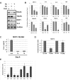
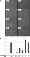
Thyroid carcinomas that harbor RET/PTC oncogenes are well differentiated, relatively benign neoplasms compared with those expressing oncogenic RAS or BRAF mutations despite signaling through shared transforming pathways. A distinction, however, is that RET/PTCs induce immunostimulatory programs, suggesting that, in the case of this tumor type, the additional pro-inflammatory pathway reduces aggressiveness. Here, we demonstrate that pro-inflammatory programs are selectively activated by TRAF2 and TRAF6 association with RET/PTC oncoproteins. Eliminating this mechanism reduces pro-inflammatory cytokine production without decreasing transformation efficiency. Conversely, ablating MEK/ERK or PI3K/AKT signaling eliminates transformation but not pro-inflammatory cytokine secretion. Functional uncoupling of the two pathways demonstrates that intrinsic pro-inflammatory pathways are not required for cellular transformation and suggests a need for further investigation into the role inflammation plays in thyroid tumor progression.
Figures


Similar articles
-
XB130, a tissue-specific adaptor protein that couples the RET/PTC oncogenic kinase to PI 3-kinase pathway.Oncogene. 2009 Feb 19;28(7):937-49. doi: 10.1038/onc.2008.447. Epub 2008 Dec 8. Oncogene. 2009. PMID: 19060924
-
BRAF mediates RET/PTC-induced mitogen-activated protein kinase activation in thyroid cells: functional support for requirement of the RET/PTC-RAS-BRAF pathway in papillary thyroid carcinogenesis.Endocrinology. 2006 Feb;147(2):1014-9. doi: 10.1210/en.2005-0280. Epub 2005 Oct 27. Endocrinology. 2006. PMID: 16254036
-
RET/PTC (rearranged in transformation/papillary thyroid carcinomas) tyrosine kinase phosphorylates and activates phosphoinositide-dependent kinase 1 (PDK1): an alternative phosphatidylinositol 3-kinase-independent pathway to activate PDK1.Mol Endocrinol. 2003 Jul;17(7):1382-94. doi: 10.1210/me.2002-0402. Epub 2003 May 8. Mol Endocrinol. 2003. PMID: 12738763
-
Genetic alterations in the phosphatidylinositol-3 kinase/Akt pathway in thyroid cancer.Thyroid. 2010 Jul;20(7):697-706. doi: 10.1089/thy.2010.1646. Thyroid. 2010. PMID: 20578891 Free PMC article. Review.
-
Treatment directed to signalling molecules in patients with advanced differentiated thyroid cancer.Anticancer Agents Med Chem. 2013 Mar;13(3):483-95. doi: 10.2174/1871520611313030011. Anticancer Agents Med Chem. 2013. PMID: 22583421 Review.
Cited by
-
TRAF6 regulates proliferation, apoptosis, and invasion of osteosarcoma cell.Mol Cell Biochem. 2012 Dec;371(1-2):177-86. doi: 10.1007/s11010-012-1434-4. Epub 2012 Aug 12. Mol Cell Biochem. 2012. PMID: 22886393
-
Exploring the Mechanism of Baicalin Intervention in Breast Cancer Based on MicroRNA Microarrays and Bioinformatics Strategies.Evid Based Complement Alternat Med. 2021 Dec 20;2021:7624415. doi: 10.1155/2021/7624415. eCollection 2021. Evid Based Complement Alternat Med. 2021. PMID: 34966436 Free PMC article.
-
miRNA-200c inhibits invasion and metastasis of human non-small cell lung cancer by directly targeting ubiquitin specific peptidase 25.Mol Cancer. 2014 Jul 6;13:166. doi: 10.1186/1476-4598-13-166. Mol Cancer. 2014. PMID: 24997798 Free PMC article.
-
The circular RNA circBCL2L1 regulates innate immune responses via microRNA-mediated downregulation of TRAF6 in teleost fish.J Biol Chem. 2021 Oct;297(4):101199. doi: 10.1016/j.jbc.2021.101199. Epub 2021 Sep 15. J Biol Chem. 2021. PMID: 34536420 Free PMC article.
-
Intracellular signal transduction and modification of the tumor microenvironment induced by RET/PTCs in papillary thyroid carcinoma.Front Endocrinol (Lausanne). 2012 May 22;3:67. doi: 10.3389/fendo.2012.00067. eCollection 2012. Front Endocrinol (Lausanne). 2012. PMID: 22661970 Free PMC article.
References
-
- Bozec A., Lassalle S., Hofman V., Ilie M., Santini J., Hofman P. (2010) The thyroid gland. A crossroad in inflammation-induced carcinoma? An ongoing debate with new therapeutic potential. Curr. Med. Chem. 17, 3449–3461 - PubMed
-
- Guarino V., Castellone M. D., Avilla E., Melillo R. M. (2010) Thyroid cancer and inflammation. Mol. Cell. Endocrinol. 321, 94–102 - PubMed
-
- Ulrich C. M., Bigler J., Potter J. D. (2006) Nonsteroidal anti-inflammatory drugs for cancer prevention. Promise, perils, and pharmacogenetics. Nat. Rev. Cancer 6, 130–140 - PubMed
Publication types
MeSH terms
Substances
Grants and funding
LinkOut - more resources
Full Text Sources
Medical
Research Materials
Miscellaneous

