. 2012 Feb 10;287(7):4451-61.
doi: 10.1074/jbc.M111.278325. Epub 2011 Dec 9.

Hydrogen sulfide inhibits high glucose-induced matrix protein synthesis by activating AMP-activated protein kinase in renal epithelial cells

Affiliations

Hydrogen sulfide inhibits high glucose-induced matrix protein synthesis by activating AMP-activated protein kinase in renal epithelial cells

Hak Joo Lee et al. J Biol Chem. .

Abstract

Hydrogen sulfide, a signaling gas, affects several cell functions. We hypothesized that hydrogen sulfide modulates high glucose (30 mm) stimulation of matrix protein synthesis in glomerular epithelial cells. High glucose stimulation of global protein synthesis, cellular hypertrophy, and matrix laminin and type IV collagen content was inhibited by sodium hydrosulfide (NaHS), an H(2)S donor. High glucose activation of mammalian target of rapamycin (mTOR) complex 1 (mTORC1), shown by phosphorylation of p70S6 kinase and 4E-BP1, was inhibited by NaHS. High glucose stimulated mTORC1 to promote key events in the initiation and elongation phases of mRNA translation: binding of eIF4A to eIF4G, reduction in PDCD4 expression and inhibition of its binding to eIF4A, eEF2 kinase phosphorylation, and dephosphorylation of eEF2; these events were inhibited by NaHS. The role of AMP-activated protein kinase (AMPK), an inhibitor of protein synthesis, was examined. NaHS dose-dependently stimulated AMPK phosphorylation and restored AMPK phosphorylation reduced by high glucose. Compound C, an AMPK inhibitor, abolished NaHS modulation of high glucose effect on events in mRNA translation as well as global and matrix protein synthesis. NaHS induction of AMPK phosphorylation was inhibited by siRNA for calmodulin kinase kinase β, but not LKB1, upstream kinases for AMPK; STO-609, a calmodulin kinase kinase β inhibitor, had the same effect. Renal cortical content of cystathionine β-synthase and cystathionine γ-lyase, hydrogen sulfide-generating enzymes, was significantly reduced in mice with type 1 diabetes or type 2 diabetes, coinciding with renal hypertrophy and matrix accumulation. Hydrogen sulfide is a newly identified modulator of protein synthesis in the kidney, and reduction in its generation may contribute to kidney injury in diabetes.


Figures
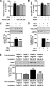
FIGURE 1.
FIGURE 1.
Hydrogen sulfide inhibits high glucose-stimulated protein synthesis, cellular hypertrophy, and matrix protein expression. Cells were quiesced in serum-free medium for 24 h followed by treatment with 100 or 250 μm NaHS with or without 30 mm glucose (high Glu) for 48 h. Control cells were grown in normal growth medium containing 5 mm glucose. A, de novo protein synthesis was measured by [35S]methionine incorporation into TCA-precipitable protein. Composite data from four experiments are shown in the graph (**, p < 0.01 versus control, #, p < 0.05 versus Glu by ANOVA). B, cellular hypertrophy was calculated as total cellular protein per unit cell number. Following treatment of GECs with high glucose for 48 h with or without NaHS, cells were lifted by trypsinization and equally divided into two tubes. Total cell number was counted in one tube, and the other tube was used for total protein estimation. Composite data from four experiments are shown in the graph (***, p < 0.001 versus control, ###, p < 0.001 versus Glu by ANOVA). C and D, GECs were treated as mentioned above, and equal amounts of cell lysate protein were separated by SDS-PAGE and immunoblotted with laminin γ1 (Lamγ1) and Col IV α5 antibodies. Loading was assessed by immunoblotting for actin. A representative blot and composite graph from three experiments are shown (*, p < 0.05; ***, p < 0.001 versus control, #, p < 0.05; ###, p < 0.001 versus Glu by ANOVA). E, cells were quiesced in serum-free medium for 24 h followed by treatment with vehicle or 250 μm NaHS with 5 or 30 mm glucose (G) for 30 min. After 30 min, the media were changed with fresh media without NaHS with respective glucose concentrations. Following incubation for 48 h, equal amounts of cell lysate protein were separated by SDS-PAGE and immunoblotted with laminin γ1 antibody. Loading was assessed by immunoblotting for actin. A representative blot and composite graph from three experiments are shown (***, p < 0.001 versus 5 mm glucose + vehicle, ###, p < 0.001 versus 30 mm glucose + vehicle by ANOVA).
FIGURE 2.
FIGURE 2.
Hydrogen sulfide inhibits high glucose-stimulated initiation phase of mRNA translation. Quiescent cells were incubated with 30 mm Glu with or without 250 μm NaHS for 30 and 60 min. Equal amounts of cell lysate protein were immunoblotted with specific antibodies. A, antibody against phosphorylated Thr-36/47 on 4E-BP1 (P-4E-BP1) or 4E-BP1. B, antibody against phosphorylated Thr-389 on p70S6 kinase (P-S6K) or p70S6 kinase (S6K). Composite data from three experiments are shown as a histogram (*, p < 0.05; **, p < 0.01 versus control; #, p < 0.05 versus Glu by ANOVA). C, antibody against PDCD4 or actin. Composite data from eight experiments are shown as a histogram (*, p < 0.05 versus control; ###, p < 0.001 versus Glu by ANOVA). D, antibody against PDCD4 or phosphorylated p70S6 kinase or p70S6 kinase or hemagglutinin (HA). Composite data from four experiments are shown as a histogram (*, p < 0.05; ***, p < 0.001 versus control; ###, p < 0.001 versus Glu by ANOVA). KD S6K, kinase-dead S6K. E, immunoprecipitation (IP) was done on equal amounts of cell lysate proteins with antibody against eIF4A followed by immunoblotting (IB) with antibody against eIF4G or PDCD4. Composite data from four experiments are shown as a histogram (*, p < 0.05 versus control; #, p < 0.05 versus Glu by ANOVA). F, experiments were done as described above with or without incubation for 1 h with 10 μm MG-132, a proteasomal inhibitor. Equal amounts of cell lysates were immunoblotted with antibody against PDCD4 and actin. A representative blot and composite graph from four experiments is shown. *, p < 0.05 versus control; #, p < 0.05 versus Glu by ANOVA).
FIGURE 3.
FIGURE 3.
Hydrogen sulfide inhibits high glucose-stimulated elongation phase of mRNA translation. Quiescent cells were incubated with 30 mm Glu with or without 250 μm NaHS for 30 and 60 min. Equal amounts of cell lysate protein were immunoblotted with specific antibodies. A, antibody against phosphorylated eEF2 kinase on Ser-366 (P-eEF2K) and eEF2 kinase (eEF2K). B, antibody against phosphorylated eEF2 on Thr-56 (P-eEF2) and eEF2. A representative blot and composite graphs from four experiments are shown (*, p < 0.05 versus control; #, p < 0.05 versus Glu by ANOVA).
FIGURE 4.
FIGURE 4.
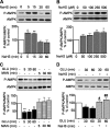
Hydrogen sulfide restores high glucose-induced reduction in AMPK phosphorylation. A, quiescent cells were treated with 250 μm NaHS for the indicated times. Equal amounts of cell lysate protein were immunoblotted with antibody against α subunit of AMPK phosphorylated on Thr-172 (P-AMPK) or AMPK antibody. A representative blot and composite graph from four experiments are shown (*, p < 0.05; **, p < 0.01 versus control by ANOVA). B, quiescent cells were treated with the indicated concentration of NaHS for 5 min. A representative blot and composite graph from three experiments are shown (*, p < 0.05 versus control by ANOVA). C, quiescent cells were incubated with 30 mm Glu or 5 mm glucose + 25 mm mannitol for the indicated times. A representative blot and composite graph from five experiments are shown (*, p < 0.05; **, p < 0.01 versus control by ANOVA). D, quiescent cells were incubated with 30 mm glucose with or without 250 μm NaHS for 30 and 60 min. Equal amounts of cell lysate protein were immunoblotted with antibody against phosphorylated Thr-172 on the α subunit of AMPK or AMPK antibody. A representative blot and composite graph from four experiments are shown (*, p < 0.05 versus control, #, p < 0.05; ##, p < 0.01 versus Glu by ANOVA).
FIGURE 5.
FIGURE 5.
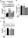
AMPK mediates hydrogen sulfide inhibition of high glucose-induced protein synthesis and matrix laminin increment. Quiescent cells were preincubated with 10 μm Compound C (CC) for 30 min. Cells were then incubated with 30 mm Glu with or without 250 μm NaHS for 30 and 60 min for studies on AMPK phosphorylation and for 48 h for evaluation of protein synthesis. A, changes in phosphorylation of AMPK (P-AMPK) or AMPK were detected by immunoblotting using the respective antibodies. A representative blot and composite graph from seven experiments are shown (*, p < 0.05 versus control, #, p < 0.05; ##, p < 0.01 versus Glu, §, p < 0.05 versus Glu+NaHS by ANOVA). B, de novo protein synthesis was measured by incorporation with [35S]methionine into TCA-precipitable protein as described under “Experimental Procedures.” Composite data from four experiments are shown in the graph (***, p < 0.001 versus control, ##, p < 0.01 versus Glu, §, p < 0.05 versus Glu+NaHS by ANOVA). C, laminin γ1 content was detected by immunoblotting against the laminin γ1 antibody. A representative blot and composite graph from three experiments are shown (**, p < 0.01 versus control, ##, p < 0.01 versus Glu, §, p < 0.05 versus Glu+NaHS by ANOVA).
FIGURE 6.
FIGURE 6.
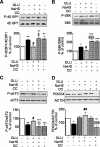
AMPK mediates hydrogen sulfide inhibition of high glucose-stimulated mTOR complex 1 activation. Quiescent cells were preincubated with 10 μm Compound C (CC) for 30 min. Cells were incubated with high glucose with or without 250 μm NaHS for 30 and 60 min. Equal amounts of cell lysate protein were immunoblotted with antibody against the indicated proteins. A, antibody against phospo-4E-BP1 (P-4E-BP1) (Thr-37/46) or 4E-BP1 (**, p < 0.01 versus control, #, p < 0.05 versus Glu; §, p < 0.05 versus Glu+NaHS by ANOVA). Representative blots and histograms on composite data from five experiments are shown. B, antibody against phospho-p70S6 kinase (P-S6K) (Thr-389) and p70S6 kinase (S6K) (*, p < 0.05; ***, p < 0.001 versus control, #, p < 0.05 versus Glu, §§§, p < 0.001 versus Glu+NaHS by ANOVA). Representative blots and histograms on composite data from five experiments are shown. C, antibody against phospho-eEF2 (p-eEF2) (Thr-56) or eEF2 (**, p < 0.01; ***, p < 0.001 versus control, #, p < 0.05 versus Glu, §§, p < 0.01 versus Glu+NaHS by ANOVA). Representative blots and histograms on composite data from five experiments are shown. D, antibody against PDCD4 or actin (*, p < 0.05; **, p < 0.01 versus control, ##, p < 0.01 versus Glu, §§§, p < 0.001 versus Glu+NaHS by ANOVA). Representative blots and histograms on composite data from four experiments are shown.
FIGURE 7.
FIGURE 7.
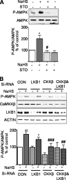
Hydrogen sulfide increases AMPK phosphorylation through the CaMKKβ pathway. A, serum-starved GECs were preincubated with 10 μm STO-609 (STO), a selective inhibitor of CaMKKβ, for 1 h followed by treatment with or without 250 μm NaHS for 5 min. Equal amounts of cell lysate protein were immunoblotted with antibody against phosphorylated Thr-172 on the α subunit of AMPK (p-AMPK) or AMPK antibody. A representative blot and composite graph from four experiments are shown (*, p < 0.05 versus control, #, p < 0.05 versus NaHS by ANOVA). B, control (CON) scrambled RNA and siRNA for LKB1 and/or CaMKKβ (CKKβ) was diluted into the siRNA transfection medium to a final concentration of 2–20 nm. Diluted siRNA was incubated with 6 μl of siRNA transfection reagent for 30 min at room temperature. GECs were washed with PBS twice and then incubated with the siRNA transfection medium for 30 min. After 30 min, cells were incubated with the diluted siRNA of control and LKB1 or CaMKKβ for 8 h, and then medium was changed to growth medium for 48 h. After 48 h, GECs were quiesced in serum-free medium for 24 h. GECs were treated with 250 μm NaHS for 5 min. Equal amounts of cell lysates were immunoblotted with the indicated antibodies. The histogram represents the composite data from five experiments (*, p < 0.05; **, p < 0.01 versus control, ###, p < 0.001 versus NaHS alone by ANOVA).
FIGURE 8.
FIGURE 8.
Expression of hydrogen sulfide-generating enzymes is reduced in the kidney in diabetes. A and B, expression of cystathionine β-synthase (CBS) and cystathionine γ-lyase (CSE) is reduced in the renal cortex of mice with type 1 diabetes (A) or type 2 (B) diabetes. Equal amounts of tissue homogenate protein were immunoblotted with antibody against CBS or cystathionine γ-lyase; loading was assessed by immunoblotting for actin (*, p < 0.05; **, p < 0.01 versus control by t test). C, immunoperoxidase staining of the kidney showed reduction in the expression of cystathionine β-synthase in type 1 diabetic mice (OVE26) when compared with non-diabetic NJ control mice.
FIGURE 9.
FIGURE 9.
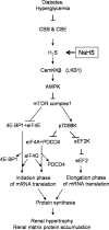
Schematic showing signaling pathways involved in hydrogen sulfide amelioration of high glucose-induced protein synthesis. CBS, cystathionine β-synthase; CSE, cystathionine γ-lyase.

Similar articles

Cited by

References

    1. Scotland R. S., Madhani M., Chauhan S., Moncada S., Andresen J., Nilsson H., Hobbs A. J., Ahluwalia A. (2005) Investigation of vascular responses in endothelial nitric-oxide synthase/cyclooxygenase-1 double-knockout mice: key role for endothelium-derived hyperpolarizing factor in the regulation of blood pressure in vivo. Circulation 111, 796–803 - PubMed
    1. Abe K., Kimura H. (1996) The possible role of hydrogen sulfide as an endogenous neuromodulator. J. Neurosci. 16, 1066–1071 - PMC - PubMed
    1. Yang G., Wu L., Jiang B., Yang W., Qi J., Cao K., Meng Q., Mustafa A. K., Mu W., Zhang S., Snyder S. H., Wang R. (2008) H2S as a physiologic vasorelaxant: hypertension in mice with deletion of cystathionine γ-lyase. Science 322, 587–590 - PMC - PubMed
    1. Zhao W., Wang R. (2002) H2S-induced vasorelaxation and underlying cellular and molecular mechanisms. Am. J. Physiol. Heart Circ. Physiol. 283, H474–H480 - PubMed
    1. Elrod J. W., Calvert J. W., Morrison J., Doeller J. E., Kraus D. W., Tao L., Jiao X., Scalia R., Kiss L., Szabo C., Kimura H., Chow C. W., Lefer D. J. (2007) Hydrogen sulfide attenuates myocardial ischemia-reperfusion injury by preservation of mitochondrial function. Proc. Natl. Acad. Sci. U.S.A. 104, 15560–15565 - PMC - PubMed

Publication types

MeSH terms

Substances