. 2012 Feb 3;110(3):471-80.
doi: 10.1161/CIRCRESAHA.111.258871. Epub 2011 Dec 8.

H2O2-induced dilation in human coronary arterioles: role of protein kinase G dimerization and large-conductance Ca2+-activated K+ channel activation

Affiliations

H2O2-induced dilation in human coronary arterioles: role of protein kinase G dimerization and large-conductance Ca2+-activated K+ channel activation

David X Zhang et al. Circ Res. .

Abstract

Rationale: Hydrogen peroxide (H(2)O(2)) serves as a key endothelium-derived hyperpolarizing factor mediating flow-induced dilation in human coronary arterioles (HCAs). The precise mechanisms by which H(2)O(2) elicits smooth muscle hyperpolarization are not well understood. An important mode of action of H(2)O(2) involves the oxidation of cysteine residues in its target proteins, including protein kinase G (PKG)-Iα, thereby modulating their activities.

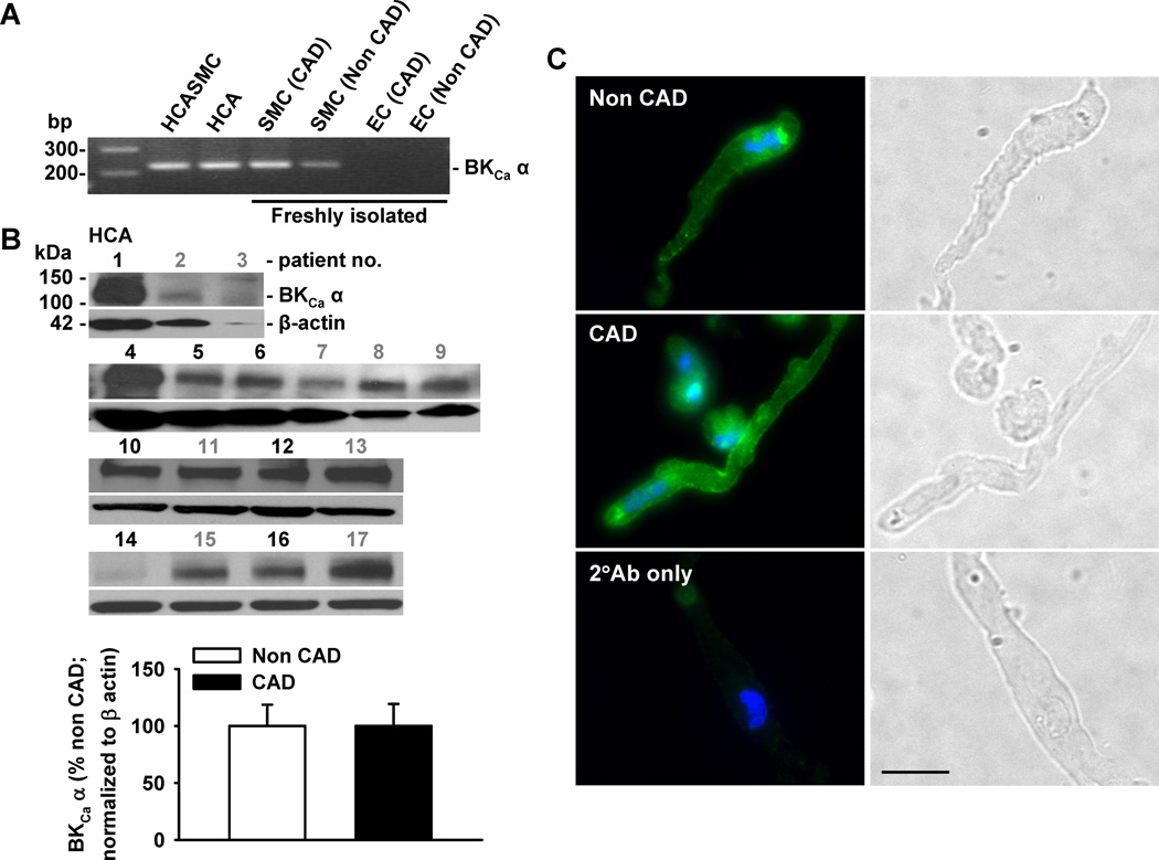
Objective: Here we hypothesize that H(2)O(2) dilates HCAs through direct oxidation and activation of PKG-Iα leading to the opening of the large-conductance Ca(2+)-activated K(+) (BK(Ca)) channel and subsequent smooth muscle hyperpolarization.

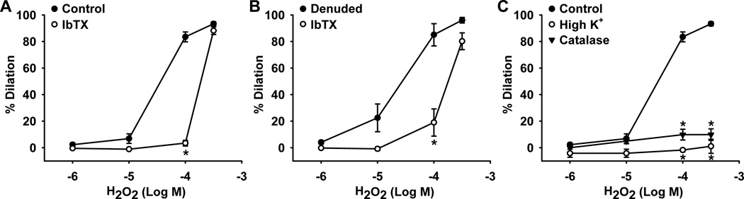
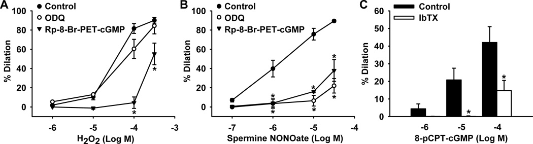
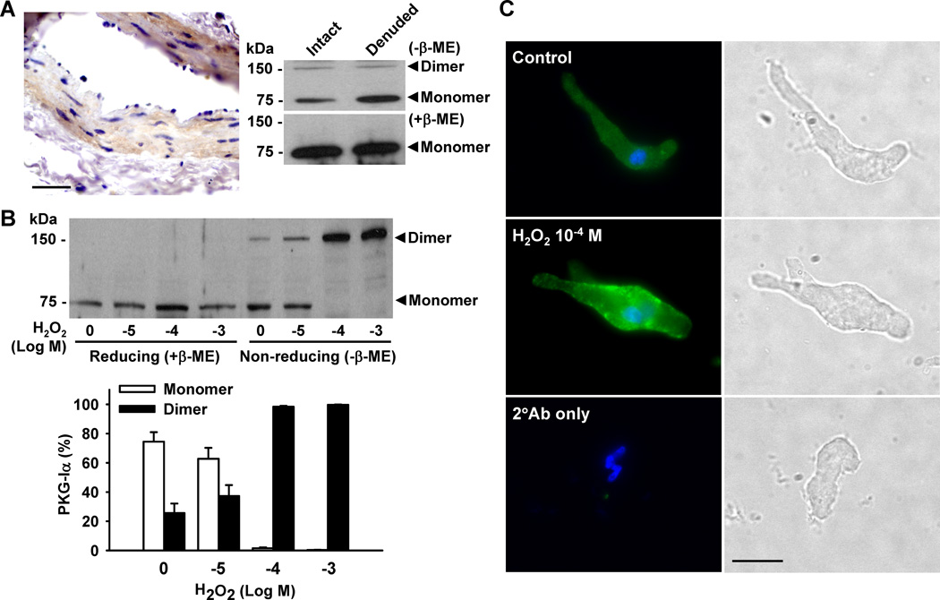
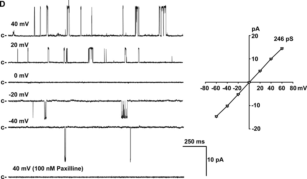
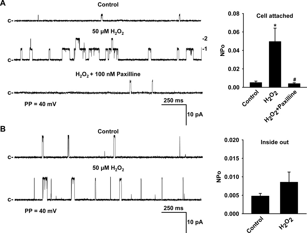
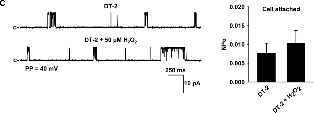
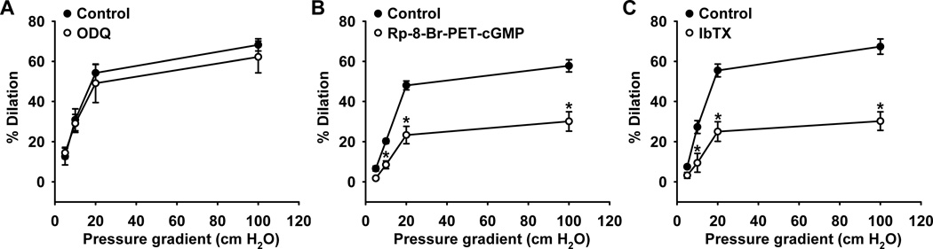
Methods and results: Flow and H(2)O(2) induced pressure gradient/concentration-dependent vasodilation in isolated endothelium-intact and -denuded HCAs, respectively. The dilation was largely abolished by iberiotoxin, a BK(Ca) channel blocker. The PKG inhibitor Rp-8-Br-PET-cGMP also markedly inhibited flow- and H(2)O(2)-induced dilation, whereas the soluble guanylate cyclase inhibitor ODQ had no effect. Treatment of coronary smooth muscle cells (SMCs) with H(2)O(2) elicited dose-dependent, reversible dimerization of PKG-Iα, and induced its translocation to the plasma membrane. Patch-clamp analysis identified a paxilline-sensitive single-channel K(+) current with a unitary conductance of 246-pS in freshly isolated coronary SMCs. Addition of H(2)O(2) into the bath solution significantly increased the probability of BK(Ca) single-channel openings recorded from cell-attached patches, an effect that was blocked by the PKG-Iα inhibitor DT-2. H(2)O(2) exhibited an attenuated stimulatory effect on BK(Ca) channel open probability in inside-out membrane patches.

Conclusions: H(2)O(2) dilates HCAs through a novel mechanism involving protein dimerization and activation of PKG-Iα and subsequent opening of smooth muscle BK(Ca) channels.


Figures

Figure 1
Figure 1. Role of BKCa channels in H2O2-mediated dilation of human coronary arterioles
H2O2 induced similar dose-dependent dilation of both endothelium-intact (A) and -denuded (B) arterioles. These relaxations were inhibited by 100 nmol/L iberiotoxin (IbTX), a BKCa channel blocker, and eliminated by high K+ (80 mmol/L) and exogenous catalase (1,000 U/ml) (C). n=3–13 vessels/each group; *P<0.05 vs. control.
Figure 2
Figure 2. Role of guanylate cyclase and PKG in H2O2- and NO-mediated relaxation of human coronary arterioles
A: H2O2-induced relaxation responses were inhibited by the PKG inhibitor Rp-8-Br-PET-cGMP (100 µmol/L) but not by the soluble guanylate cyclase inhibitor ODQ (10 µmol/L). B: The NO donor spermine NONOate-induced relaxations that were inhibited by both Rp-8-Br-PET-cGMP and ODQ. C: The stable cGMP analogue 8-pCPT-cGMP dose-dependently dilated coronary arterioles, a response inhibited by the BKCa channel blocker iberiotoxin (100 nmol/L). n=4–15 vessels/each group; *P<0.05 vs. control.
Figure 3
Figure 3. H2O2-induced protein dimerization of PKG-I in human coronary artery smooth muscle cells
A: The protein expression of PKG-I in HCAs was detected by immunohistochemical analysis (left panel; scale bar = 50 µm) and Western blot (right panel). Under basal conditions, PKG-I was primarily in monomeric form. B: Treatment of HCASMCs with H2O2 induced concentration-dependent dimerization of PKG-I which was blocked by the reducing agent β-mercaptoethanol (β-ME). Data are representative of 3 independent experiments. C: Immunofluorescence detected diffuse cytosolic expression of PKG-I under control conditions (upper). H2O2 (100 µmol/L) induced punctate expression of PKG-I along the plasma membrane (middle). Scale bar = 20 µm. Data are representative of 3 independent experiments with 5–10 cells/group/experiment.
Figure 4
Figure 4. Expression of BKCa channels in coronary smooth muscle cells
A: Expression of BKCa channel α-subunits was detected at mRNA level in freshly isolated SMCs from patients with and without CAD, as indicated by a representative image of RT-PCR analysis. B: BKCa α-subunit protein was expressed in coronary arterioles from patients with (patient no. in black) and without CAD (patient no. in gray). Lower, summarized data; n=8 and 9 for CAD and non CAD, respectively. C: Presence of BKCa channel α-subunit protein (green) is confirmed with immunocytochemistry using freshly dispersed SMCs. Cell nuclei are stained in blue. Scale bar = 20 µm. Data are representative of 3 independent experiments with 5–10 cells/ group/ experiment. D: In inside-out patches of freshly isolated SMCs from human coronary arterioles, an increase in membrane potential enhanced BKCa channel open probability (left) that was abolished by 100 nmol/L paxilline, a specific BKCa channel inhibitor. The current-voltage relationship revealed a unitary conductance of 246 pS with a reversal potential of 0 mV in symmetrical (145 mmol/L) K+ solutions (right). c, closed state. n=4 patches.
Figure 5
Figure 5. Effect of H2O2 on BKCa channel currents in freshly isolated coronary arteriolar smooth muscle cells
Using cell-attached patches (A), H2O2 (50 µmol/L) increased BKCa single-channel currents in a paxilline (100 nmol/L)-sensitive fashion. However, H2O2-induced BKCa activation was greatly diminished in single-channel recordings from inside-out patches which lack intracellular constituents (B). H2O2-activated BKCa single-channel currents recorded from cell-attached patches were reduced in the presence of 10 µmol/L DT-2, a PKG-Iα inhibitor (C). c, closed state; PP, patch potential. n=6–12 patches/each group; *P<0.05 vs. control, #P<0.05 vs. H2O2.
Figure 6
Figure 6. Role of guanylate cyclase, PKG, and BKCa channels in human coronary arteriolar relaxation to flow
Fluid flow induced dilation of human coronary arterioles was attenuated by the PKG inhibitor Rp-8-Br-PET-cGMP (B) and by the BKCa channel blocker iberiotoxin (C). The guanylate cyclase inhibitor ODQ, had no effect (A). n=3-6 vessels/each group; *P<0.05 vs. control.

References

    1. Perez-Vizcaino F, Cogolludo A, Moreno L. Reactive oxygen species signaling in pulmonary vascular smooth muscle. Respir Physiol Neurobiol. 2010;174:212–220. - PubMed
    1. Schröder E, Eaton P. Hydrogen peroxide as an endogenous mediator and exogenous tool in cardiovascular research: issues and considerations. Curr Opin Pharmacol. 2008;8:153–159. - PubMed
    1. Zhang DX, Gutterman DD. Mitochondrial reactive oxygen species-mediated signaling in endothelial cells. Am J Physiol Heart Circ Physiol. 2007;292:H2023–H2031. - PubMed
    1. Liu Y, Bubolz AH, Mendoza S, Zhang DX, Gutterman DD. H2O2 is the transferrable factor mediating flow-induced dilation in human coronary arterioles. Circ Res. 2011;108:566–573. - PMC - PubMed
    1. Larsen BT, Gutterman DD, Sato A, Toyama K, Campbell WB, Zeldin DC, Manthati VL, Falck JR, Miura H. Hydrogen peroxide inhibits cytochrome p450 epoxygenases: interaction between two endothelium-derived hyperpolarizing factors. Circ Res. 2008;102:59–67. - PMC - PubMed

Publication types

MeSH terms