. 2011 Dec 7;3(112):112ra122.
doi: 10.1126/scitranslmed.3002761.

Age-related intimal stiffening enhances endothelial permeability and leukocyte transmigration

Affiliations

Age-related intimal stiffening enhances endothelial permeability and leukocyte transmigration

John Huynh et al. Sci Transl Med. .

Abstract

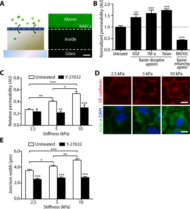
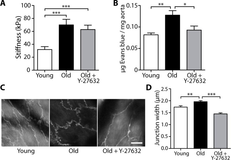
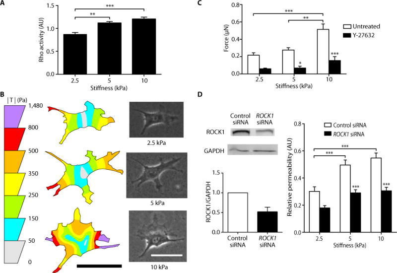
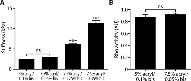
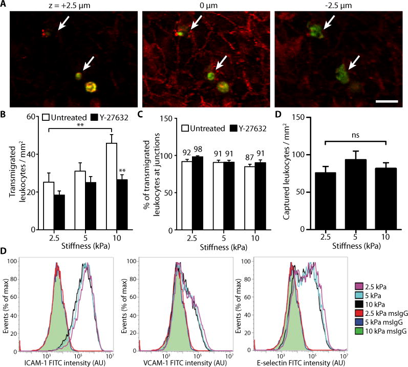
Age is the most significant risk factor for atherosclerosis; however, the link between age and atherosclerosis is poorly understood. During both aging and atherosclerosis progression, the blood vessel wall stiffens owing to alterations in the extracellular matrix. Using in vitro and ex vivo models of vessel wall stiffness and aging, we show that stiffening of extracellular matrix within the intima promotes endothelial cell permeability--a hallmark of atherogenesis. When cultured on hydrogels fabricated to match the elasticity of young and aging intima, endothelial monolayers exhibit increased permeability and disrupted cell-cell junctions on stiffer matrices. In parallel experiments, we showed a corresponding increase in cell-cell junction width with age in ex vivo aortas from young (10 weeks) and old (21 to 25 months) healthy mice. To investigate the mechanism by which matrix stiffening alters monolayer integrity, we found that cell contractility increases with increased matrix stiffness, mechanically destabilizing cell-cell junctions. This increase in endothelial permeability results in increased leukocyte extravasation, which is a critical step in atherosclerotic plaque formation. Mild inhibition of Rho-dependent cell contractility using Y-27632, an inhibitor of Rho-associated kinase, or small interfering RNA restored monolayer integrity in vitro and in vivo. Our results suggest that extracellular matrix stiffening alone, which occurs during aging, can lead to endothelial monolayer disruption and atherosclerosis pathogenesis. Because previous therapeutics designed to decrease vascular stiffness have been met with limited success, our findings could be the basis for the design of therapeutics that target the Rho-dependent cellular contractile response to matrix stiffening, rather than stiffness itself, to more effectively prevent atherosclerosis progression.


Conflict of interest statement

Competing interests: The authors declare no competing interests.

Figures

Fig. 1
Fig. 1
Matrix stiffness increases endothelial permeability and cell-cell junction width. (A) A cartoon shows FITC-dextran permeation through an endothelial monolayer into a polyacrylamide gel. On the right is a representative confocal cross-section of dextran capture in a 2.5-kPa polyacrylamide gel, depicting the method used to measure permeability. Dashed lines represent the top and bottom edges of the gel. Permeability values of cell (BAEC)-seeded gels were calculated as the pixel intensity within the gel divided by the pixel intensity above the gel normalized to that calculated in cell-free gels. Scale bar, 20 μm. (B) Effects of known barrier-disruptive (VEGF, TNF-α, or thrombin) and barrier-enhancing (BW245C) agonists on the permeability of BAECs cultured on 5-kPa gels. Permeability values are normalized to that of untreated cells. Data are means ± SEM. **P < 0.01, ***P < 0.001 (Dunnett’s test) compared to untreated cells. (C) Relative permeability of BAECs cultured on substrates matching (2.5 kPa) and exceeding (5 and 10 kPa) the stiffness of bovine arterial intima with (n = 8 gels, three independent experiments) or without (n = 16–20 gels, ten independent experiments) Y-27632 treatment. Data are means ± SEM. *P < 0.05, **P < 0.01, ***P < 0.001 (Tukey’s test) compared to respective untreated conditions unless otherwise indicated by brackets. (D) Fluorescent images showing VE-cadherin (red), actin (green), and nuclei (blue) of confluent BAECs on gels. (E) VE-cadherin junction-width measurements of BAECs on gels with or without 30 min Y-27632 treatment (n = 60 junction measurements, two independent experiments). Scale bar, 10 μm. Data are means ± SEM. *P < 0.05, **P < 0.01, ***P < 0.001 (Tukey’s test) compared to respective untreated conditions unless otherwise indicated by brackets.
Fig. 2
Fig. 2
Aging increases subendothelial stiffness and endothelial intercellular junction separation in mice. (A) AFM indentation measurements of thoracic aorta of untreated young (10 to 11 weeks, n = 61 indent sites, 3 aortas) and old (20 to 25 months, n = 55 indent sites, 5 aortas) mice, as well as Y-27632-treated old (20 months, n = 76 indent sites, 4 aortas) mice. Data are means ± SEM. ***P < 0.001 (Dunn’s test). (B) Permeability measurements of thoracic aorta of untreated young (9 weeks, n = 4 aortas) and old (19 to 22 months, n = 6 aortas) mice, as well as Y-27632-treated old (20 months, n = 4 aortas) mice. Data are means ± SEM. *P < 0.05, **P < 0.01 (Dunn’s test). (C) Two-photon microscopy of VE-cadherin immunofluorescent staining in endothelial cells of intact, untreated young (10 week) and old (16 month) thoracic mouse aortas, as well as Y-27632-treated old (24 month) thoracic aortas. Scale bar, 10 μm. (D) VE-cadherin junction width measurements of thoracic aortic endothelial cells from untreated young (10 weeks, n = 240 width measurements, 4 aortas) and old (16 to 24 months, n = 390 width measurements, 7 aortas) mice, as well as Y-27632-treated old (21 to 24 months, n = 156 width measurements, 3 aortas) mice. Data are means ± SEM. **P < 0.01, ***P < 0.001 (Dunn’s test).
Fig. 3
Fig. 3
Matrix stiffness increases Rho activity and cell contractility, and inhibition of cell contractility restores barrier integrity. (A) Rho activity of BAECs cultured on polyacrylamide gels of different stiffness (three independent experiments, each performed in duplicate). Data are means ± SEM. **P < 0.01, ***P < 0.001 (Tukey’s test). (B) Representative traction force distribution maps and phase images of BAECs on gels. Scale bars, 50 μm. (C) Traction force microscopy measurements of cell contractility with (n = 9–12 cells) or without (n = 19–34 cells) Y-27632 treatment. Data are means ± SEM. *P < 0.05, **P < 0.01, ***P < 0.001 (Tukey’s test) compared to respective untreated conditions, unless otherwise indicated by brackets. (D) RNAi of ROCK1 decreases stiffness-induced endothelial permeability. (Left) Western blot analysis and densitometry quantification of RNAi of ROCK1 (n = 2). (Right) Relative permeability of BAECs cultured on gels of varying stiffnesses with (n = 13–15 gels) or without (n = 13 gels) siRNA knockdown of ROCK1. Data are means ± SEM. *P < 0.05, **P < 0.01, ***P < 0.001 (Tukey’s test) compared to respective control siRNA, unless otherwise indicated by brackets.
Fig. 4
Fig. 4
Gels made from different acrylamide concentrations that are similar in stiffness elicit similar levels of Rho activation. (A) Stiffness measurements of polyacrylamide gels of varying percentages of acrylamide and bisacrylamide. Data are means ± SEM (n = 7–8 measurements, 2 gels per group). ***P < 0.001, ns (not significant), (Tukey’s test) compared to 5% acrylamide/0.1% bisacrylamide. (B) Intracellular Rho activity of bovine aortic endothelial cells seeded on polyacrylamide gels of similar stiffness. Data for the 5% acrylamide/0.1% bisacrylamide is re-plotted from the 2.5 kPa group of Fig. 3A. Data are means ± SEM (n = 3 independent experiments, each performed in duplicate). ns, not significant (Student’s t test).
Fig. 5
Fig. 5
Matrix stiffness enhances leukocyte transmigration. (A) Representative fluorescent images of leukocytes (green) transmigrating through an endothelial monolayer (VE-cadherin, red). Images were taken at focal planes above the monolayer (+2.5 μm), at the monolayer (0 μm), and below the monolayer (−2.5 μm). Arrows indicate transmigrated cells. Scale bar, 20 μm. (B) Histogram showing the effect of matrix stiffness on the number of transmigrated leukocytes. Endothelial cells were either untreated (n = 19 fields of view, three independent experiments) or pre-treated with Y-27632 (n = 17–18 fields of view). Data are means ± SEM. **P < 0.01 (Tukey’s test) compared to respective untreated condition, unless otherwise indicated by brackets. (C) Percentage of leukocytes that transmigrated through intercellular junctions. Numbers above bars indicate the nominal values. Data are means ± SEM. (D) The effect of substrate stiffness on the number of captured leukocytes. Data are means ± SEM (n = 79 to 96 fields of view per stiffness). ns, not significant (Tukey’s test). (E) Flow cytometry analysis of HUVEC expression of ICAM-1, VCAM-1, and E-selectin on gels after a 6-h treatment with TNF-α. Shaded green curves represent overlapping mouse IgG isotype controls (msIgG) for each of the three matrices. Three independent experiments were performed. No statistically significant differences (Tukey’s test) were found between the three stiffnesses for all three adhesion molecules.

Comment in

  • ROCK in a stiff place.
    Lauffenburger DA. Lauffenburger DA. Sci Transl Med. 2011 Dec 7;3(112):112fs12. doi: 10.1126/scitranslmed.3003389. Sci Transl Med. 2011. PMID: 22158859

References

    1. Laurent S, Boutouyrie P. Recent advances in arterial stiffness and wave reflection in human hypertension. Hypertension. 2007;49:1202–1206. - PubMed
    1. Fernandes VR, Polak JF, Cheng S, Rosen BD, Carvalho B, Nasir K, McClelland R, Hundley G, Pearson G, O’Leary DH, Bluemke DA, Lima JA. Arterial stiffness is associated with regional ventricular systolic and diastolic dysfunction: the Multi-Ethnic Study of Atherosclerosis. Arterioscler Thromb Vasc Biol. 2008;28:194–201. - PubMed
    1. McEniery CM, Yasmin, Hall IR, Qasem A, Wilkinson IB, Cockcroft JR. Normal vascular aging: differential effects on wave reflection and aortic pulse wave velocity: the Anglo-Cardiff Collaborative Trial (ACCT) J Am Coll Cardiol. 2005;46:1753–1760. - PubMed
    1. Zieman SJ, Melenovsky V, Kass DA. Mechanisms, pathophysiology, and therapy of arterial stiffness. Arterioscler Thromb Vasc Biol. 2005;25:932–943. - PubMed
    1. Greenwald SE. Ageing of the conduit arteries. J Pathol. 2007;211:157–172. - PubMed

Publication types