. 2012 Feb 3;287(6):4198-210.
doi: 10.1074/jbc.M111.296475. Epub 2011 Dec 12.

Enhanced killing of cancer cells by poly(ADP-ribose) polymerase inhibitors and topoisomerase I inhibitors reflects poisoning of both enzymes

Affiliations

Enhanced killing of cancer cells by poly(ADP-ribose) polymerase inhibitors and topoisomerase I inhibitors reflects poisoning of both enzymes

Anand G Patel et al. J Biol Chem. .

Abstract

Poly(ADP-ribose) polymerase-1 (PARP1) plays critical roles in the regulation of DNA repair. Accordingly, small molecule inhibitors of PARP are being developed as agents that could modulate the activity of genotoxic chemotherapy, such as topoisomerase I poisons. In this study we evaluated the ability of the PARP inhibitor veliparib to enhance the cytotoxicity of the topoisomerase I poisons topotecan and camptothecin (CPT). Veliparib increased the cell cycle and cytotoxic effects of topotecan in multiple cell line models. Importantly, this sensitization occurred at veliparib concentrations far below those required to substantially inhibit poly(ADP-ribose) polymer synthesis and at least an order of magnitude lower than those involved in selective killing of homologous recombination-deficient cells. Further studies demonstrated that veliparib enhanced the effects of CPT in wild-type mouse embryonic fibroblasts (MEFs) but not Parp1(-/-) MEFs, confirming that PARP1 is the critical target for this sensitization. Importantly, parental and Parp1(-/-) MEFs had indistinguishable CPT sensitivities, ruling out models in which PARP1 catalytic activity plays a role in protecting cells from topoisomerase I poisons. To the contrary, cells were sensitized to CPT in a veliparib-independent manner upon transfection with PARP1 E988K, which lacks catalytic activity, or the isolated PARP1 DNA binding domain. These results are consistent with a model in which small molecule inhibitors convert PARP1 into a protein that potentiates the effects of topoisomerase I poisons by binding to damaged DNA and preventing its normal repair.


Figures
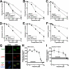
FIGURE 1.
FIGURE 1.
Veliparib enhances the antiproliferative effects of topotecan and CPT. A2780 (A), SKOV3 (B), Ovcar5 (C), and A549 (D and E) cells were exposed to topotecan (A–D) or CPT (E) for 24 h in the absence or presence of the indicated concentration of veliparib, then washed and incubated in drug-free medium until they formed colonies. F, HL-60 cells were treated for 24 h with the indicated topotecan concentration in the absence or presence of 1 μm veliparib, washed, and allowed to form colonies in soft agar in the absence of drug. Error bars, ± S.D. of triplicate (A–E) or quadruplicate (F) plates from the assays shown. Experiments in each panel are representative of three independent assays. G, shown is localization of PARP1 (green) and pADPr (red) in cells treated for 30 min with diluent or 1 mm MMS. H and I, shown is mean fluorescence of nuclei after treatment of cells with 1 mm MMS (H) or 4 μm topotecan (I) in the presence of the indicated veliparib concentration followed by fixation and staining for pADPr. Error bars, ± S.E. for 30 nuclei. Gray horizontal bars indicate signal from diluent-treated cells.
FIGURE 2.
FIGURE 2.
BRCA2-mutant hamster cells are sensitized to CPT at veliparib concentrations of that fail to induce synthetic lethality. A and C, V79 (BRCA2-positive) and V-C8 (BRCA2-negative) Chinese hamster cells were treated with CPT alone for 24 h (A) or with veliparib alone for 48 h (C), then washed and incubated in drug-free medium for 5–7 days until colonies formed. B, cells were treated with the indicated concentrations of veliparib continuously. D and E, V79 (D) or V-C8 (E) cells were incubated for 24 h with the indicated concentrations of CPT in the presence of 100 nm veliparib or diluent (0.2% DMSO), then washed and incubated in drug-free medium for 5–7 days until colonies formed. Error bars, ± S.D. of triplicate plates from the assays shown. Experiments are representative of three independent assays with each drug.
FIGURE 3.
FIGURE 3.
Effect of veliparib on topotecan-induced changes in cell cycle distribution, drug accumulation, and stabilization of Top1cc. A, A549 cells were treated for 48 h with the indicated concentration of topotecan in the absence or presence of 1 μm veliparib, fixed in ethanol, and stained with propidium iodide. B, a graphic representation of the results from panel A shows induction of G2-phase arrest in the presence of topotecan. Similar results were observed in two additional independent assays. C, flow cytometry histograms show fluorescence of cells treated with diluent or topotecan in the absence or presence of 1 μm veliparib (left) or 1 μm canertinib (right). D, topotecan uptake into the indicated cell lines in the absence or presence of 1 μm veliparib is shown. Uptake in the absence of veliparib was set to 100%. Canertinib served as a positive control for inhibition of topotecan transporters in A549 cells (66). Error bars, ± S.E. of three independent assays. E, predicted shifts in cleavage-religation equilibrium based on previous reports that PARP1 binds to and facilitates topo I-mediated resealing are depicted. In this model inhibition of PARP1 would be predicted to result in accumulation of topo I-DNA covalent complexes (Top1cc). F, alkaline elution was performed to measure protein-linked DNA single-strand breaks in A549 cells exposed for 45 min at 37 °C to the indicated topotecan concentrations in the presence or absence of 1 μm veliparib. Lower inset, shown is a radiation standard curve from this experiment. Upper inset, shown are the summarized results of four independent experiments comparing the DNA single-strand breaks detected by alkaline elution in the presence of proteinase K after treatment with 250 nm topotecan in the absence and presence of 1 μm veliparib. Gray horizontal line, mean of four determinations.
FIGURE 4.
FIGURE 4.
PARP inhibition sensitizes cells to topo I poisons, but targeted Parp1 deletion or PARP1 siRNA does not. A, shown are DNA histograms of Parp1+/+ and Parp1−/− MEFs. Numbers represent the means ± S.D. from four independent experiments. B, shown is an immunoblot of whole cell lysates from Parp1+/+ and Parp1−/− MEFs. Lamins A and C served as loading controls. C, MEFs were exposed to CPT in the presence or absence of 1 μm veliparib for 24 h, washed, and allowed to form colonies in drug-free medium. D, colony formation by a pair of 3T3 fibroblasts from independently generated Parp1 knock-out and control mice treated as in panel C is shown. Inset, shown is an immunoblot of whole cell lysates from Parp1+/+ and Parp1−/− 3T3 fibroblasts. Hsp90β served as a loading control. The asterisk indicates nonspecific band. E, shown is clonogenic survival of A2780 cells treated with PARP1 shRNA versus nontargeting control beginning 3 days before a 24-h exposure to the indicated concentration of topotecan. Inset, shown is an immunoblot of whole cell lysates harvested from a portion of the same cell pools at the initiation of topotecan exposure. Error bars, ± S.D. of triplicate aliquots. Similar results were observed in three-five independent experiments for each panel.
FIGURE 5.
FIGURE 5.
Sensitization of Parp1−/− MEFs by catalytically inactive PARP1 or its DBD. A, shown is a schematic of PARP1 constructs transfected into Parp1−/− MEFs. BRCT, BRCA1 C-terminal; NLS, nuclear localization signal. B, shown is an immunoblot of whole cell lysates from Parp1−/− MEFs transfected with the indicated construct. Hsp90β served as a loading control. C–E, MEFs transfected with empty vector or cDNA encoding wt PARP1, PARP1 E988K, or PARP1 DBD were exposed to CPT in the presence or absence of 1 μm veliparib for 24 h, washed, and allowed to form colonies in drug-free medium. Results from a single experiment have been separated into three panels for clarity. Error bars, ± S.D. of triplicate aliquots. Similar results were observed in three independent experiments for each panel.
FIGURE 6.
FIGURE 6.
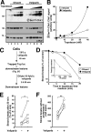
Detection of increased DNA damage in the presence of veliparib. A, after log phase A549 cells were treated for 24 h with 0, 5, 10, 20, or 40 nm topotecan in the absence or presence of 1 μm veliparib, whole cell lysates were subjected to SDS-PAGE and probed with antibodies to the indicated antigens. c-Raf served as a loading control. Numbers at the left are molecular mass markers in kDa. B, shown is a ratio of signals in the corresponding phospho-Chk1 and Chk1 lanes in panel A (arbitrary units). C, shown is a schematic of the experiment in panels D–F. A549 cells were treated with 250 nm topotecan for 45 min at 37 °C in the presence of diluent or 1 μm veliparib, diluted 10-fold into medium in the continued presence of diluent or veliparib, incubated for 2.5–20 min, and applied to filters for alkaline elution and immediately lysed in SDS. D, shown is a relative number of strand breaks persisting after the indicated incubation. Results are representative of five independent experiments. Inset, radiation calibration curve from this experiment. E, shown is the relative number of strand breaks persisting 20 min after dilution into medium containing diluent or 1 μm veliparib. *, p < 0.04 level by sign test. F, 15 min after cells were diluted 10-fold as indicated in panels C and E, the remaining strand breaks were detected by alkaline elution in the absence of proteinase K. Results shown are from four independent experiments. **, p = 0.01 by paired t test. Horizontal gray bars, mean of four experiments.

References

    1. Heck M. M., Hittelman W. N., Earnshaw W. C. (1988) Differential expression of DNA topoisomerases I and II during the eukaryotic cell cycle. Proc. Natl. Acad. Sci. U.S.A. 85, 1086–1090 - PMC - PubMed
    1. Potmesil M., Hsiang Y. H., Liu L. F., Bank B., Grossberg H., Kirschenbaum S., Forlenza T. J., Penziner A., Kanganis D., Forlenzar T. J. (1988) Resistance of human leukemic and normal lymphocytes to drug-induced DNA cleavage and low levels of DNA topoisomerase II. Cancer Res. 48, 3537–3543 - PubMed
    1. Champoux J. J. (2001) DNA topoisomerases. Structure, function, and mechanism. Annu. Rev. Biochem. 70, 369–413 - PubMed
    1. Pommier Y., Leo E., Zhang H., Marchand C. (2010) DNA topoisomerases and their poisoning by anticancer and antibacterial drugs. Chem. Biol. 17, 421–433 - PMC - PubMed
    1. Muller M. T. (1985) Quantitation of eukaryotic topoisomerase I reactivity with DNA. Preferential cleavage of supercoiled DNA. Biochim. Biophys. Acta 824, 263–267 - PubMed

Publication types

MeSH terms

LinkOut - more resources