. 2012 Feb 17;287(8):5253-66.
doi: 10.1074/jbc.M111.276162. Epub 2011 Dec 12.

Small molecule structure correctors abolish detrimental effects of apolipoprotein E4 in cultured neurons

Affiliations

Small molecule structure correctors abolish detrimental effects of apolipoprotein E4 in cultured neurons

Hung-Kai Chen et al. J Biol Chem. .

Abstract

Apolipoprotein E4 (apoE4), the major genetic risk factor for late onset Alzheimer disease, assumes a pathological conformation, intramolecular domain interaction. ApoE4 domain interaction mediates the detrimental effects of apoE4, including decreased mitochondrial cytochrome c oxidase subunit 1 levels, reduced mitochondrial motility, and reduced neurite outgrowth in vitro. Mutant apoE4 (apoE4-R61T) lacks domain interaction, behaves like apoE3, and does not cause detrimental effects. To identify small molecules that inhibit domain interaction (i.e. structure correctors) and reverse the apoE4 detrimental effects, we established a high throughput cell-based FRET primary assay that determines apoE4 domain interaction and secondary cell- and function-based assays. Screening a ChemBridge library with the FRET assay identified CB9032258 (a phthalazinone derivative), which inhibits domain interaction in neuronal cells. In secondary functional assays, CB9032258 restored mitochondrial cytochrome c oxidase subunit 1 levels and rescued impairments of mitochondrial motility and neurite outgrowth in apoE4-expressing neuronal cells. These benefits were apoE4-specific and dose-dependent. Modifying CB9032258 yielded well defined structure-activity relationships and more active compounds with enhanced potencies in the FRET assay (IC(50) of 23 and 116 nm, respectively). These compounds efficiently restored functional activities of apoE4-expressing cells in secondary assays. An EPR binding assay showed that the apoE4 structure correction resulted from direct interaction of a phthalazinone. With these data, a six-feature pharmacophore model was constructed for future drug design. Our results serve as a proof of concept that pharmacological intervention with apoE4 structure correctors negates apoE4 detrimental effects in neuronal cells and could be further developed as an Alzheimer disease therapeutic.


Figures

FIGURE 1.
FIGURE 1.
Chemical structures of representative phthalazinone analogs.
FIGURE 2.
FIGURE 2.
FRET assay used to monitor intracellular apoE4 domain interaction in Neuro-2a cells and to identify apoE4 structure correctors. A, apoE4 domain interaction allows FRET to occur between the donor and acceptor fluorophores. Inhibiting domain interaction by structure correctors reduces FRET from the apoE4 reporter. SC, structure corrector. B, FRET intensity (ratio of FRET signal divided by GFP signal) from GFP-apoE4-eDHFR is 36.3% higher than GFP-apoE3-eDHFR. *, p < 0.0001 (n = 6). FRET intensity from GFP-apoE4/eDHFR (expressing GFP-apoE4 and eDHFR by separate constructs, n = 5) is comparable with that of GFP-apoE3-eDHFR. C, CB9032258 inhibits FRET intensity from GFP-apoE4-eDHFR but not GFP-apoE3-eDHFR. *, p < 0.05, by ANOVA with Tukey's post hoc test. D, dose-response curves show relative potencies of PH-001, PH-002, CB9032258, and PH-008 to inhibit FRET intensity. Values are mean ± S.D. (error bars).
FIGURE 3.
FIGURE 3.
MtCOX1 in-cell Western assay used to monitor apoE4-specific reduction of mtCOX1 in Neuro-2a cells and prevention of this impairment by apoE4 structure correctors. A, representative images showing apoE4-specific reduction of mtCOX1 immunoreactivity. Green, anti-mtCOX1 immunofluorescence. Red, counterstain of Sapphire700 (a protein dye) and DRAQ5 (a DNA stain) for cell number normalization. A merged image of both channels is also shown. B, relative mtCOX1 levels in apoE4-expressing cells are significantly lower than in control cells or cells expressing apoE3 or apoE4-R61T. Values are mean ± S.D. (error bars) from three separate experiments. Signals from 15 wells were measured for every cell type in each experiment. *, p < 0.001 by ANOVA with Tukey's post hoc test. C, dose-dependent effects of CB9032258, PH-005, and PH-002 to restore mtCOX1 levels in apoE4-expressing cells. Shown on the left are merged images of mtCOX1 immunofluorescence (green) and Sapphire700/DRAQ5 counterstains (red) from cells treated with various doses of structure correctors. CB9032258 increases mtCOX1 levels only at doses of 3330 and 10,000 nm, whereas PH-005 is effective at lower doses (1110–10,000 nm). PH-002, the most active one, significantly increases mtCOX1 levels at a dose as low as 41.2 nm. The dose-response curves for the three compounds are shown on the right. Values are mean ± S.D. from triplicate wells. D, CB9032258 increases mtCOX1 levels only in apoE4-expressing cells and not in control cells or cells expressing apoE3 or apoE4-R61T. Values are mean ± S.D. from triplicate wells. *, p < 0.01 by ANOVA with Tukey's post hoc test. Similar results were obtained in at least three separate experiments. E, significant correlation of potencies between FRET and mtCOX1 assays. Pearson's r = 0.803, R2 = 0.645, p < 0.0001, n = 47 phthalazinone analogs.
FIGURE 4.
FIGURE 4.
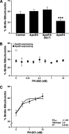
Mitochondrial motility assay used to monitor apoE4-specific reduction in percentage of motile mitochondria and the reversibility of this impairment by apoE4 structure correctors. A, differentiated PC12 cells were incubated for 24 h at 37 °C without (control) or with exogenous apoE3, apoE4, or apoE4-R61T (7.5 μg/ml). Mitochondrial dynamics, analyzed as a percentage of motile mitochondria, was measured during a 15-min recording (12 frames/min) (control, n = 218 mitochondria from 12 cells; apoE3, n = 209 mitochondria from 14 cells; apoE4, n = 212 mitochondria from nine cells; apoE4-R61T, n = 164 mitochondria from 12 cells). Values are mean ± S.E. (error bars). ***, p < 0.001 versus control (two-tailed t test). B, mitochondrial motility analyzed in apoE3- and apoE4-expressing PC12 cells at 37 °C without the addition of small molecules served as a control. After the addition of increasing doses of inactive PH-008 incubated for 24 h, the percentage of motile mitochondria was determined in the differentiated PC12 cells expressing apoE4 (n = 100 mitochondria from 10 cells in two separate experiments). Values are mean ± S.E. C, mitochondrial motility analyzed in apoE4-expressing PC12 cells at 37 °C with increasing concentrations of the active phthalazinone PH-001. Values are mean ± S.E. EC50, Hill slope, and R2 reported for the positive dose response.
FIGURE 5.
FIGURE 5.
Active phthalazinone analogs reverse apoE4-specific effects on neurite outgrowth. A, representative images show that apoE4 expression is associated with fewer Neuro-2a cells with long neurites, whereas apoE3- and apoE4-R61T-expressing cells have similarly long neurites. ApoE4-expressing cells treated with the active phthalazinone PH-002 have similar long neurites resembling the apoE4-R61T cells. B, dose-response relationship of phthalazinone analogs on neurite outgrowth in apoE4-expressing Neuro-2a cells. Values are mean ± S.D. (error bars) (n = 4). p values (no addition controls versus neurite outgrowth with increasing concentration of structure correctors) were as follows: PH-001, PH-002, and PH-004 at 3 nm, p < 0.0005; PH-001 and PH-004 at 10 nm, p < 0.0001 (PH-002, p = 0.003); PH-001 and PH-002 at 30 nm, p < 0.003 (PH-004, p < 0.0001); PH-001, PH-002, PH-004, and CB9032258, p = 0.0008, p = 0.007, p = 0.002, and p = 0.02, respectively.
FIGURE 6.
FIGURE 6.
PH-002 increases COX1 levels in primary neurons from NSE-apoE4 mouse cortex and hippocampus. PH-002 (200 nm) was incubated with apoE4-expressing primary neurons for 4 days, and then the level of COX1 was determined by Western blot (mean ± S.D. (error bars) for three replicates). VDAC1 was used as the internal standard.
FIGURE 7.
FIGURE 7.
Demonstration of direct binding of PH-002 to the apoE4 amino-terminal domain by EPR. Incubation of the apoE4 amino-terminal domain with increasing concentrations of PH-002 resulted in a dose-dependent effect on the mobility of the spin label at position Cys-76, as determined from the effect on the control peak of the EPR spectra (h1/h0). Results are mean ± S.E. (error bars); control versus PH-002/apoE at ratios of 2:1 and 4:1; p = 0.005 and p = 0.002, respectively (n = 3).
FIGURE 8.
FIGURE 8.
Pharmacophore modeling for phthalazinone analogs using a ligand-based methodology. A, phthalazinone pharmacophore. The six-feature pharmacophore is composed of three hydrophobic spheres (blue; spheres are labeledA”, “E”, and “F”) and three hydrogen bond acceptors (green arrows with arrowheads pointing toward donor labeledB”, “C”, and “D”). B, pharmacophore fitted with PH-002. This potent compound fits all six features. C, two-dimensional structure of PH-002 annotated with moieties making contact with features of the pharmacophore. D, pharmacophore fitted with PH-008. This inactive compound fits five of the six features. Hydrophobe “F” is not satisfied, suggesting the importance of this feature for activity. E, pharmacophore fitted with PH-007. This inactive compound fits five of the six features. Hydrogen bond acceptor “C” is not possible with this compound. The core ring structure is not a phthalazinone but a 2-methyl-2H-isoquinolin-1-one, which lacks acceptor functionality at this position. Note that unlike PH-008, hydrophobe “F” is satisfied. The importance of the hydrogen bond acceptor “C” is suggested by the lack of activity of this compound.
FIGURE 9.
FIGURE 9.
Structure-based pharmacophore analysis docking the six-feature pharmacophore with the crystal structure of the amino-terminal domain of apoE4. A, PH-002 docked into the 22-kDa domain of human apoE4 (Protein Data Bank entry 1GS9) utilizing the six-feature pharmacophore. Purple CPK, Arg-61, which is interacting with hydrophobe “F”. Yellow CPK, Arg-119, which is interacting with the hydrogen bond acceptor “B”. B, close-up of PH-002 docking.

References

    1. Corder E. H., Saunders A. M., Strittmatter W. J., Schmechel D. E., Gaskell P. C., Small G. W., Roses A. D., Haines J. L., Pericak-Vance M. A. (1993) Gene dose of apolipoprotein E type 4 allele and the risk of Alzheimer's disease in late onset families. Science 261, 921–923 - PubMed
    1. Strittmatter W. J., Saunders A. M., Schmechel D., Pericak-Vance M., Enghild J., Salvesen G. S., Roses A. D. (1993) Apolipoprotein E. High-avidity binding to β-amyloid and increased frequency of type 4 allele in late-onset familial Alzheimer disease. Proc. Natl. Acad. Sci. U.S.A. 90, 1977–1981 - PMC - PubMed
    1. Saunders A. M., Strittmatter W. J., Schmechel D., St George-Hyslop P. H., Pericak-Vance M. A., Joo S. H., Rosi B. L., Gusella J. F., Crapper-MacLachlan D. R., Alberts M. J., Hulette C., Crain B., Goldgaber D., Roses A. D. (1993) Association of apolipoprotein E allele ϵ4 with late-onset familial and sporadic Alzheimer's disease. Neurology 43, 1467–1472 - PubMed
    1. Farrer L. A., Cupples L. A., Haines J. L., Hyman B., Kukull W. A., Mayeux R., Myers R. H., Pericak-Vance M. A., Risch N., van Duijn C. M. (1997) Effects of age, sex, and ethnicity on the association between apolipoprotein E genotype and Alzheimer disease. A meta-analysis. JAMA 278, 1349–1356 - PubMed
    1. Mahley R. W. (1988) Apolipoprotein E. Cholesterol transport protein with expanding role in cell biology. Science 240, 622–630 - PubMed

Publication types

MeSH terms

LinkOut - more resources