. 2012 Feb;32(4):751-62.
doi: 10.1128/MCB.06255-11. Epub 2011 Dec 12.

TCERG1 regulates alternative splicing of the Bcl-x gene by modulating the rate of RNA polymerase II transcription

Affiliations

TCERG1 regulates alternative splicing of the Bcl-x gene by modulating the rate of RNA polymerase II transcription

Marta Montes et al. Mol Cell Biol. 2012 Feb.

Abstract

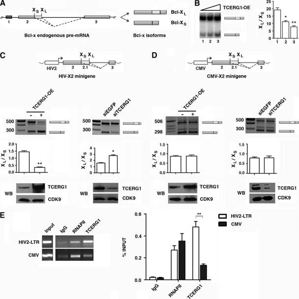
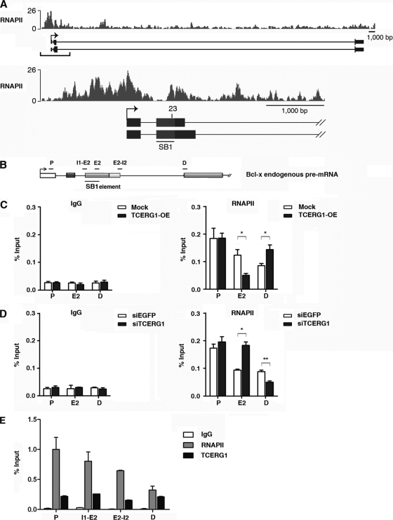
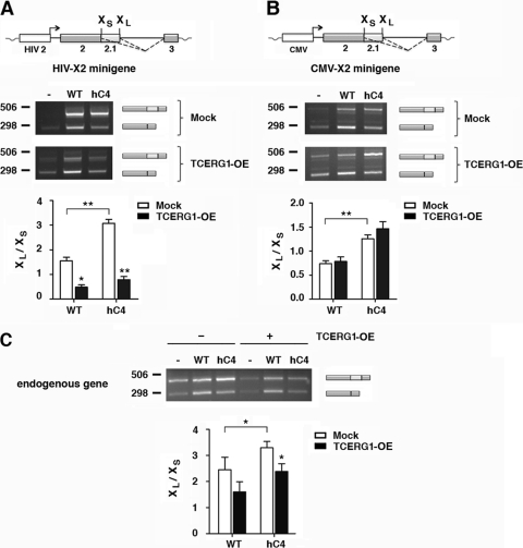
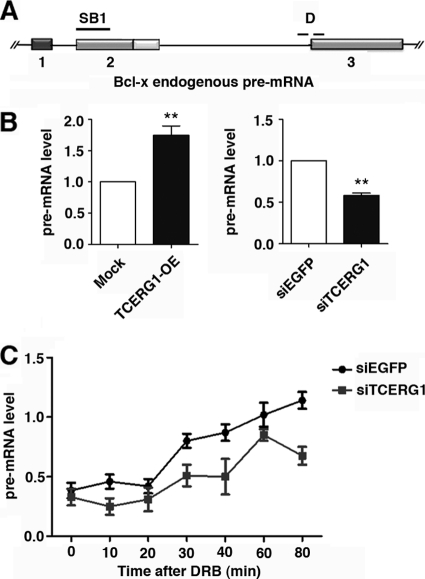
Complex functional coupling exists between transcriptional elongation and pre-mRNA alternative splicing. Pausing sites and changes in the rate of transcription by RNA polymerase II (RNAPII) may therefore have fundamental impacts in the regulation of alternative splicing. Here, we show that the elongation and splicing-related factor TCERG1 regulates alternative splicing of the apoptosis gene Bcl-x in a promoter-dependent manner. TCERG1 promotes the splicing of the short isoform of Bcl-x (Bcl-x(s)) through the SB1 regulatory element located in the first half of exon 2. Consistent with these results, we show that TCERG1 associates with the Bcl-x pre-mRNA. A transcription profile analysis revealed that the RNA sequences required for the effect of TCERG1 on Bcl-x alternative splicing coincide with a putative polymerase pause site. Furthermore, TCERG1 modifies the impact of a slow polymerase on Bcl-x alternative splicing. In support of a role for an elongation mechanism in the transcriptional control of Bcl-x alternative splicing, we found that TCERG1 modifies the amount of pre-mRNAs generated at distal regions of the endogenous Bcl-x. Most importantly, TCERG1 affects the rate of RNAPII transcription of endogenous human Bcl-x. We propose that TCERG1 modulates the elongation rate of RNAPII to relieve pausing, thereby activating the proapoptotic Bcl-x(S) 5' splice site.


Figures

Fig 1
Fig 1
Regulation of Bcl-x alternative splicing by TCERG1. (A) Schematic representation of the structure of the Bcl-x gene, with exons (boxes) and introns (lines). Two splice variants derived from the Bcl-x gene, antiapoptotic Bcl-xL and proapoptotic Bcl-xs, were generated via alternative 5′ splice site selection within exon 2. The positions of the 5′ splice sites of Bcl-xs and Bcl-xL are indicated. The dotted lines indicate the alternative splicing events. (B) Overexpression (OE) of TCERG1 increased the levels of proapoptotic Bcl-xs in a dose-dependent manner. HEK293T cells were transfected with an empty vector (lane 1) or plasmids carrying TCERG1 (lanes 2 and 3, respectively). After total RNA extraction, the RNA splicing variants were amplified by radioactive RT-PCR, and the products were separated on a native 4% polyacrylamide gel. The graph on the left shows the densitometric analyses results as the ratio of Bcl-xL to Bcl-xS isoforms from three independent experiments (means ± standard deviations [SD]). *, P < 0.05; **, P < 0.01. (C and D) Effects of TCERG1 overexpression/knockdown on alternative splicing of Bcl-x minigenes carrying the HIV-2 LTR (C) and CMV (D) promoters. A schematic of the minigenes is shown. HEK293T cells were cotransfected with the Bcl-x minigene together with empty plasmid (-) or a TCERG1 expression plasmid (+). For the RNAi experiments, HEK293T cells were cotransfected with the Bcl-x minigene together with siTCERG1 or the control (siEGFP). RT-PCR was performed to analyze alternatively spliced forms of Bcl-x. The graphs show the densitometric analysis results as the ratio of Bcl-xL to Bcl-xS isoforms from four independent experiments (means ± SD). *, P < 0.05; **, P < 0.01. A fraction of the cell lysates was analyzed by immunoblotting with the indicated antibodies to detect the TCERG1 and CDK9 proteins. (E) ChIP analysis of the recruitment of TCERG1 to the HIV-2 and CMV promoters. TCERG1 binds preferentially to the HIV-2 promoter in vivo. HEK293T cells were cotransfected with either HIV2-X2 or CMV-X2 minigenes together with a TCERG1 expression plasmid. The cross-linked and shared chromatin was immunoprecipitated with the indicated antibodies. After reversal of the cross-linking and purification of the DNA, PCR was used to detect the sequences corresponding to the HIV-2 and CMV promoters. The input shows the signal from the chromatin before immunoprecipitation. The primers used in the ChIP assays are described in Materials and Methods. The bar graph presents the quantification of the data by qPCR from four independent experiments (means ± SD). **, P < 0.01.
Fig 2
Fig 2
Effects of TCERG1 on Bcl-x splicing depend on the SB1 regulatory element. (A) Structure of the Bcl-x gene and X2 and X2.13 (ΔSB1) minigenes. (B and C) Analysis of the effects of SB1 deletion on Bcl-x splice site selection in response to TCERG1 overexpression (OE) (B) and knockdown (C). HEK293T cells were cotransfected with Bcl-X2 or Bcl-X2.13 together with empty vector (-) or TCERG1 expression plasmid. For the RNAi experiments, HEK293T cells were cotransfected with the Bcl-x minigene together with siTCERG1 or control (siEGFP). RT-PCR was performed to analyze the alternatively spliced forms of Bcl-x. The graphs show the densitometric analysis results as the ratio of Bcl-xL to Bcl-xS isoforms from three independent experiments (means ± standard deviations [SD]). *, P < 0.05; **, P < 0.01. A fraction of the cell lysates was analyzed by immunoblotting with the indicated antibodies to detect the TCERG1 and CDK9 proteins. (D) RNA coimmunoprecipitation of the Bcl-x pre-mRNA with antibodies against TCERG1 is dependent on the SB1 region. The X2 or X2.13 transcripts were incubated in HeLa nuclear extracts under splicing conditions and then immunoprecipitated using the indicated antibodies. After five washes, the RNA was precipitated and quantified. The data are presented as the percentage of the bound input (means ± SD). **, P < 0.01. (E) TCERG1 associates with transcripts derived from the endogenous Bcl-x gene. In vivo immunoprecipitation assays were carried out with specific antibodies against TCERG1. Transcripts derived from the endogenous Bcl-x gene were detected by qPCR (see Materials and Methods). The data are presented as percentages of the bound input (means ± SD). **, P < 0.01. (F) Diagrammatic representation of the deleted sequences (in bold) in the Bcl-x minigene tested in the experiments. The numbers below the lines indicate the deleted regions. (G) HEK293T cells were cotransfected with 0.5 μg HIV-2 reporter minigene (X2, wild type; X2.13, carrying a complete deletion of the SB1 element; Δ9, Δ11, Δ13, Δ16, Δ17, Δ23, carrying 10-nucleotide deletions in the SB1 element) together with 0.5 μg empty vector (-) or 0.5 μg TCERG1 expression vector (+). RT-PCR was performed to analyze the alternatively spliced forms of Bcl-x. The bar graph shows the densitometric analysis results as the ratio of Bcl-xL to Bcl-xS isoforms from three independent experiments. (H) HEK293T cells were cotransfected with 0.5 μg of the indicated Bcl-x minigenes together with siRNAs against enhanced green fluorescent protein (siEGFP) or TCERG1 (siTCERG1). RT-PCR was performed to determine alternatively spliced forms of Bcl-x. The bar graph shows the densitometric analysis results as the ratio of Bcl-xL to Bcl-xS isoforms from three independent experiments. (I) RNA coimmunoprecipitation was carried out using the Δ12 and Δ23 transcripts as described in the legend for panel D. Data are presented as percentages of the bound input (means ± SD). *, P < 0.05; **, P < 0.01.
Fig 3
Fig 3
TCERG1 modulates RNAPII distribution on Bcl-x exon 2. (A) An RNAPII-paused region coincided with region 23 in the Bcl-x gene. The densities of sequence reads from the RNAPII chromatin-immunopurified samples (bars) are displayed above the Bcl-x promoter region (−1,000 bp upstream of the start site) and the first 4,000 bp of the transcribed region. The alternative splicing regulatory region SB1 (gray box) and region 23, required for TCERG1 activity, are indicated. (B) Schematic representation of the structure of the Bcl-x gene, drawn with exons (boxes) and introns (lines). The positions of the SB1 element and of the primers used to amplify mRNA products by qPCR are indicated (P, promoter region; E2, exon 2; I1-E2, intron 1-exon 2 junction; E2-I2, exon 2-intron 2 junction; D, distal region). (C) The polymerase distribution at different positions of the gene was detected by ChIP followed by qPCR of cells transfected with an empty vector (mock) or a TCERG1 overexpression (OE) expression vector. (D) The same experiment described for panel B was carried out with siRNAs against enhanced green fluorescent protein (EGFP) or TCERG1. (E) Distributions of RNAPII and TCERG1 at different positions of the gene were detected by ChIP followed by qPCR of cells. IgG was used as a control in all the ChIP experiments. Data from three independent experiments are presented as percentages of the bound input (means ± standard deviations). *, P < 0.05; **, P < 0.01.
Fig 4
Fig 4
A slow polymerase (hC4) favors the use of the Bcl-xL splice site, and overexpression of TCERG1 promotes activation of the Bcl-xS splice site. (A) The HIV-X2 minigene was cotransfected into HEK293T cells with empty vector (-) or plasmids containing the α-amanitin-resistant wild-type (WT) or slow (hC4) RNAPII genes. In each case, the cells were also transfected with either the empty vector (mock) or the TCERG1 expression vector and treated with α-amanitin. The alternative splicing was assessed by RT-PCR as described in Materials and Methods. The data are presented as the ratios of Bcl-xL to Bcl-xS from three independent experiments (means ± standard deviations [SD]). *, P < 0.05; **, P < 0.01. (B) The same experiment described for panel A was carried out with the CMV-X2 minigene. The data are presented as the ratio of Bcl-xL to Bcl-xS from three independent experiments (means ± SD). **, P < 0.01. (C) The same experiment described for panel A was carried out in the absence of minigenes. The effect on the Bcl-x endogenous gene was assessed by RT-PCR as described in Materials and Methods. The data are presented as the ratio of Bcl-xL to Bcl-xS from three independent experiments (means ± SD). *, P = 0.0399 (WT-mock versus hC4-mock); P = 0.038 for hC4-mock versus hC4-TCERG1; P = 0.054 for WT-mock versus WT-TCERG1.
Fig 5
Fig 5
TCERG1 increases the rate of RNAPII transcription in vivo. (A) Schematic representation of the structure of the Bcl-x gene, drawn with exons (boxes) and introns (lines). The position of the SB1 element and of the primers spanning the intron-exon 3 junction (D) used to amplify transcripts by qRT-PCR are indicated. (B) Quantitative analysis of the amount of nascent Bcl-x transcripts. (Left) HEK293T cells were transfected with an empty vector (mock) or the TCERG1 overexpression (OE) plasmid. (Right) HEK293T cells were transfected with siRNAs against EGFP or TCERG1. The quantification of the experimental data from four independent experiments performed in triplicates is shown in graphic form (means ± standard deviations [SD]). **, P < 0.01. (C) Kinetics of RNAPII-dependent transcription elongation in the presence or absence of TCERG1. HEK293T cells were transfected with siEGFP or siTCERG1 and treated 48 h later with 100 μM DRB for 3 h. After DRB removal, fresh medium was added and samples were taken at the times indicated. qRT-PCR was performed using primer set D to measure the levels of pre-mRNA expression. The graph shows pre-mRNA levels at different times from the control (siEGFP) or TCERG1-depleted cells (siTCERG1). The data shown are the averages from triplicates of two independent experiments (means ± SD). *, P < 0.05; **, P < 0.01.

Similar articles

Cited by

References

    1. Alexander RD, Innocente SA, Barrass JD, Beggs JD. 2010. Splicing-dependent RNA polymerase pausing in yeast. Mol. Cell 40:582–593 - PMC - PubMed
    1. Allo M, et al. 2009. Control of alternative splicing through siRNA-mediated transcriptional gene silencing. Nat. Struct. Mol. Biol. 16:717–724 - PubMed
    1. Boise LH, et al. 1993. bcl-x, a bcl-2-related gene that functions as a dominant regulator of apoptotic cell death. Cell 74:597–608 - PubMed
    1. Bourquin JP, et al. 1997. A serine/arginine-rich nuclear matrix cyclophilin interacts with the C-terminal domain of RNA polymerase II. Nucleic Acids Res. 25:2055–2061 - PMC - PubMed
    1. Caceres JF, Kornblihtt AR. 2002. Alternative splicing: multiple control mechanisms and involvement in human disease. Trends Genet. 18:186–193 - PubMed

Publication types

Associated data