

Characterization of limbal stem cell deficiency by in vivo laser scanning confocal microscopy: a microstructural approach
- PMID: 22159172
- PMCID: PMC3928362
- DOI: 10.1001/archophthalmol.2011.378
Characterization of limbal stem cell deficiency by in vivo laser scanning confocal microscopy: a microstructural approach
Abstract
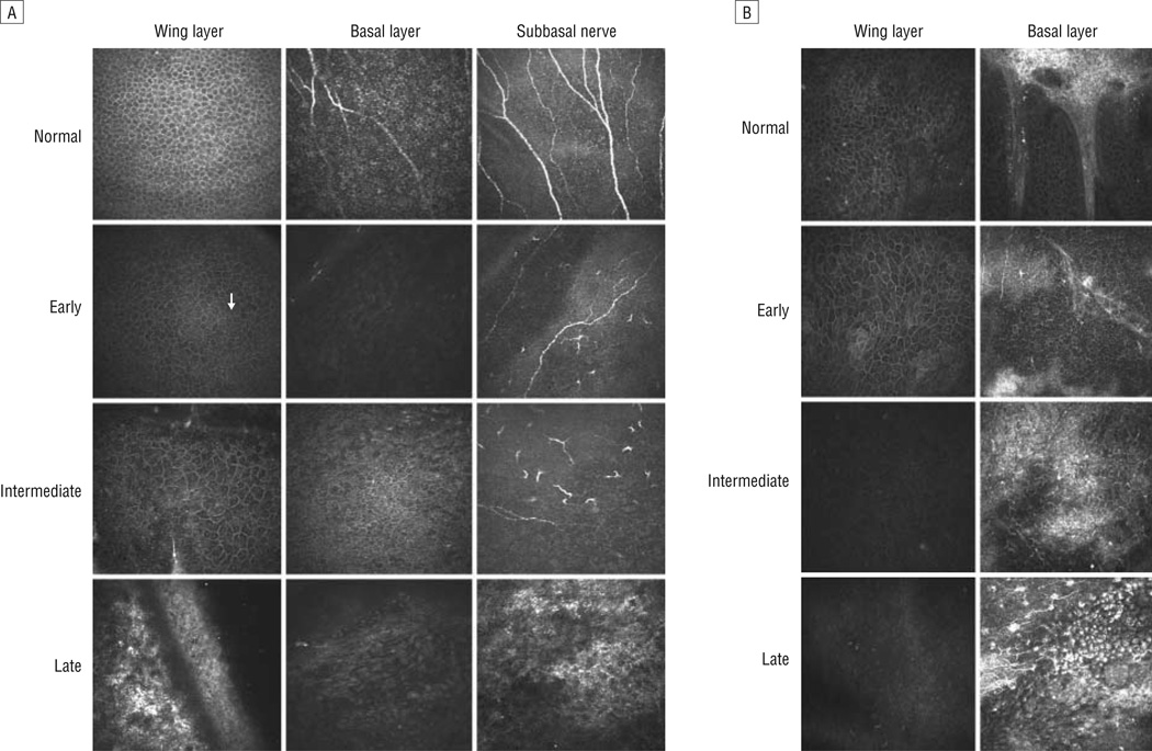
Objective: To evaluate the cellular changes in the corneal epithelium and surrounding structures in limbal stem cell deficiency (LSCD) by using in vivo laser scanning confocal microscopy.
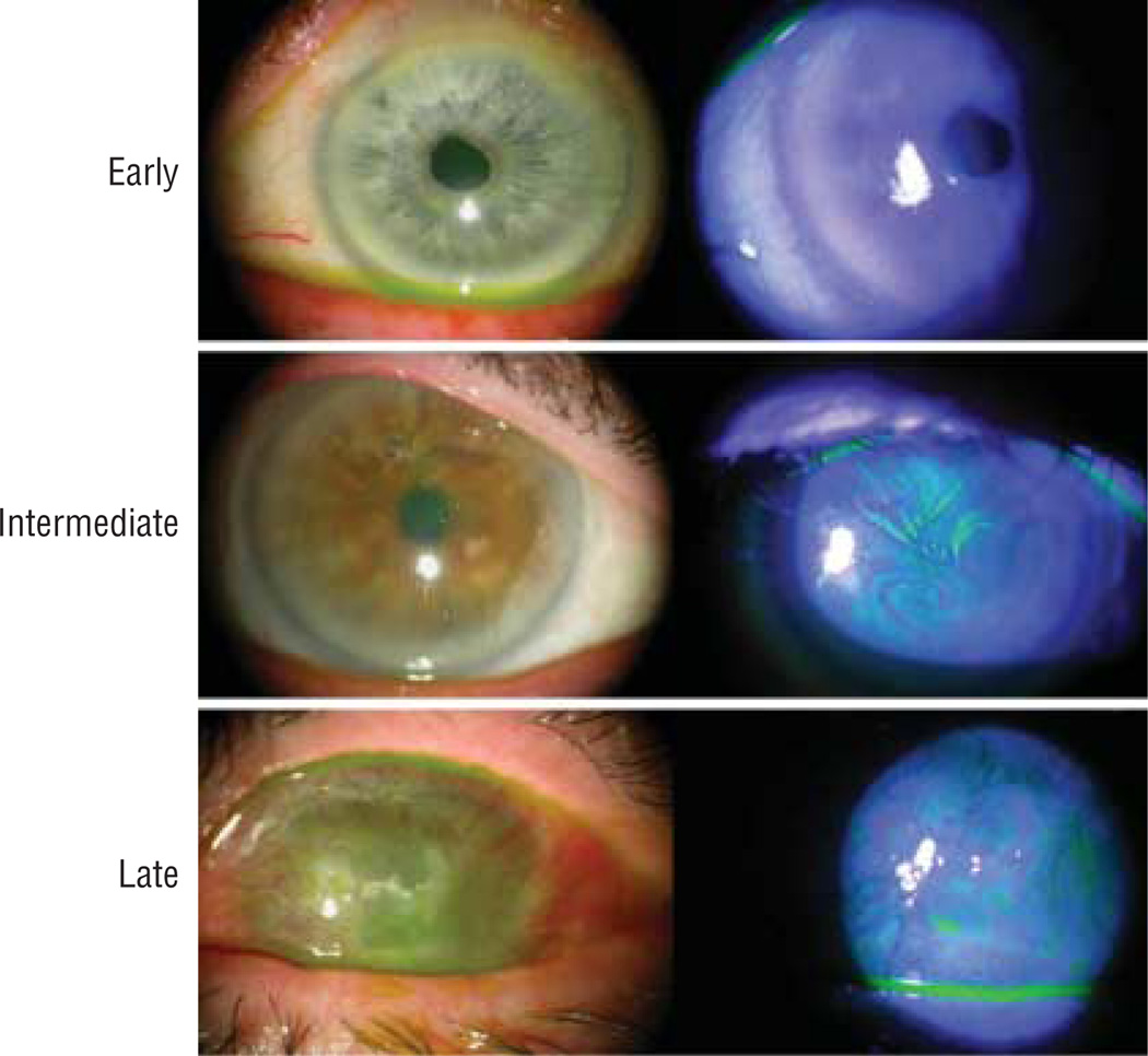
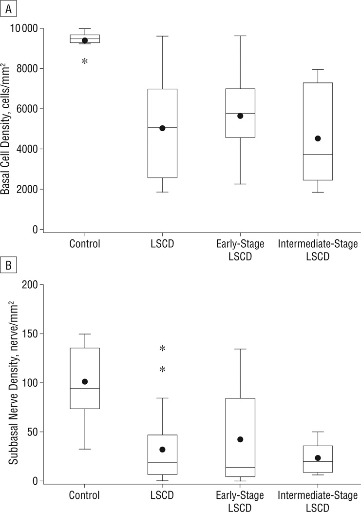
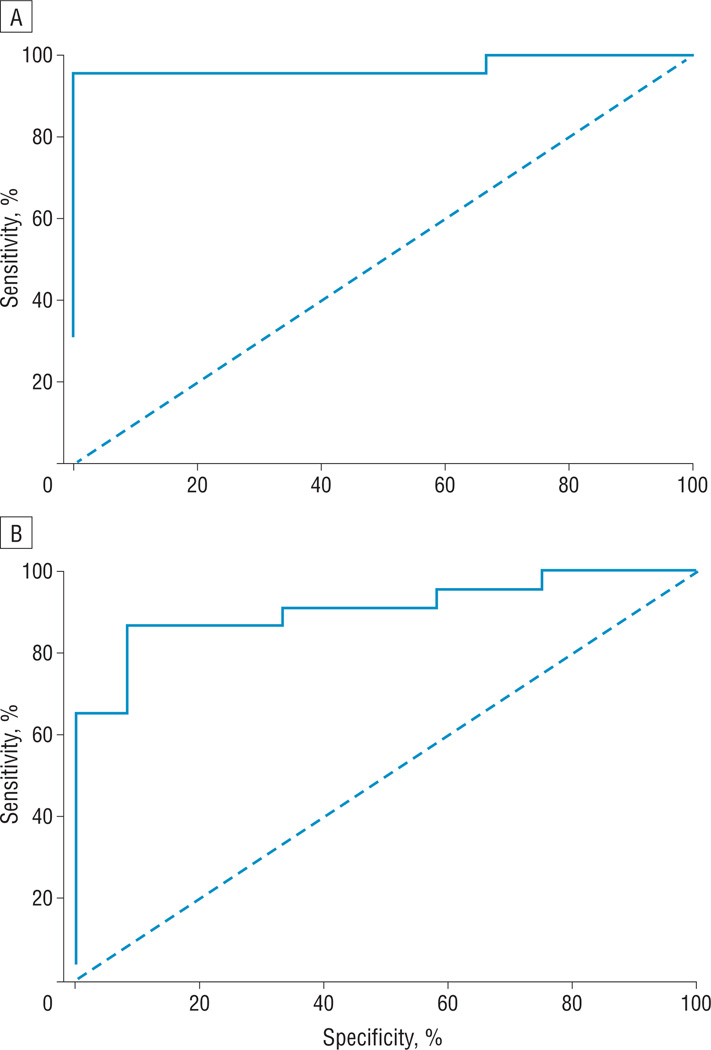
Methods: This was a prospective comparative study that included 27 eyes of 20 patients with LSCD and 12 eyes of 10 healthy subjects. All subjects underwent slitlamp examination, and LSCD was classified into 3 groups on the basis of clinical presentation. Confocal imaging of the central cornea and 4 locations of limbus was performed. Morphologic characteristics of the corneal epithelium were studied. The basal epithelial cell density and subbasal nerve density in the central cornea were calculated, and a potential correlation between the decrease in basal epithelial cell density and subbasal nerve density in LSCD was investigated.
Results: The wing and basal epithelial cells became progressively metaplastic, and the basal epithelial cell density and subbasal nerve density in the early and intermittent stages decreased significantly compared with controls (all P < .01). Normal basal epithelial cell morphology was completely lost and subbasal nerves were absent in the late stage of LSCD. The decrease in basal cell density correlated with the decrease in subbasal nerve density in patients with LSCD (P = .03).
Conclusions: There are significant microstructural changes associated with early LSCD. These cellular changes could help to understand the disease process and classify and monitor limbal stem cell dysfunction.
Figures
References
-
- Lavker RM, Tseng SC, Sun TT. Corneal epithelial stem cells at the limbus: looking at some old problems from a new angle. Exp Eye Res. 2004;78(3):433–446. - PubMed
-
- Tseng SC. Concept and application of limbal stem cells. Eye (Lond) 1989;3(pt 2):141–157. - PubMed
-
- Puangsricharern V, Tseng SC. Cytologic evidence of corneal diseases with limbal stem cell deficiency. Ophthalmology. 1995;102(10):1476–1485. - PubMed
-
- Dua HS, Azuara-Blanco A. Limbal stem cells of the corneal epithelium. Surv Ophthalmol. 2000;44(5):415–425. - PubMed
Publication types
MeSH terms
Grants and funding
LinkOut - more resources
Full Text Sources
Medical
