. 2011 Dec 1;124(Pt 23):4039-50.
doi: 10.1242/jcs.087858. Epub 2011 Dec 8.

MT1-MMP regulates the turnover and endocytosis of extracellular matrix fibronectin

Affiliations

MT1-MMP regulates the turnover and endocytosis of extracellular matrix fibronectin

Feng Shi et al. J Cell Sci. .

Abstract

The extracellular matrix (ECM) is dynamically remodeled by cells during development, normal tissue homeostasis and in a variety of disease processes. We previously showed that fibronectin is an important regulator of ECM remodeling. The deposition and/or polymerization of fibronectin into the ECM controls the deposition and stability of other ECM molecules. In addition, agents that inhibit fibronectin polymerization promote the turnover of fibronectin fibrils and enhance ECM fibronectin endocytosis and intracellular degradation. Endocytosis of ECM fibronectin is regulated by β1 integrins, including α5β1 integrin. We have examined the role of extracellular proteases in regulating ECM fibronectin turnover. Our data show that membrane type matrix metalloproteinase 1 (MT1-MMP; also known as MMP14) is a crucial regulator of fibronectin turnover. Cells lacking MT1-MMP show reduced turnover and endocytosis of ECM fibronectin. MT1-MMP regulates ECM fibronectin remodeling by promoting extracellular cleavage of fibronectin and by regulating α5β1-integrin endocytosis. Our data also show that fibronectin polymerization stabilizes fibronectin fibrils and inhibits ECM fibronectin endocytosis by inhibiting α5β1-integrin endocytosis. These data are the first to show that an ECM protein and its modifying enzyme can regulate integrin endocytosis. These data also show that integrin trafficking plays a major role in modulating ECM fibronectin remodeling. The dual dependence of ECM fibronectin turnover on extracellular proteolysis and endocytosis highlights the complex regulatory mechanisms that control ECM remodeling to ensure maintenance of proper tissue function.


Figures
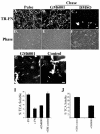
Fig. 1.
Fig. 1.
GM6001, an MMP inhibitor, prevents ECM fibronectin turnover, endocytosis and degradation. (A–F) GM6001 prevents ECM fibronectin turnover. FN-null MFs were incubated with 10 μg/ml TR–fibronectin overnight (A, Pulse). Cells were washed and then incubated for 27 hours in medium lacking fibronectin, but containing 20 μM GM6001 (B) or vehicle control (C). Scale bar: 50 μm. Corresponding phase-contrast images are shown in D–F. (G,H) GM6001 inhibits ECM fibronectin endocytosis. FN-null MFs were seeded onto pre-assembled matrix containing TR–fibronectin and cultured for 1 hour. The medium was then supplemented with 20 μM GM6001 (G) or control compound (H), and the cells cultured for an additional 36 hours. Arrows point to intracellular TR–fibronectin. Scale bar: 10 μm. (I,J) GM6001 inhibits intracellular degradation of ECM fibronectin. FN-null MFs (I) or rat SMCs (J) were incubated overnight with 125I-fibronectin. Cells were washed and then incubated with culture medium lacking or containing 10 μg/ml unlabeled fibronectin (I), 20 μM GM6001 or control compound (I,J) for 24 hours. The counts in the TCA-soluble fraction of the medium were used to determine intracellular degradation of ECM fibronectin, as described in the Materials and Methods (means ± s.d., n=3).
Fig. 2.
Fig. 2.
Membrane-type MMPs are involved in endocytosis and turnover of ECM fibronectin. FN-null MFs were seeded onto pre-assembled matrix containing TR–fibronectin (TR-FN; A–I) or Alexa-Fluor-488–FN (J) and cultured for 1–2 hours. The medium was then supplemented with 50 nM TIMP-1 (B,E,H), 50 nM TIMP-2 (C,F,I), or PBS (A,D,G), and the cells cultured for 24 hours. Cells were then fixed for imaging assay (A–I) or processed for flow cytometry analysis (J). Images in A–C were taken at low magnification and at a focal plane that would best depict fibronectin fibrils. High magnification views are shown in G–I to allow intracellular vesicles to be distinguished from bright fibrils. Arrows point to intracellular TR–fibronectin, and arrowheads point to areas that are enlarged in the insets. Scale bars: 50 μm (A–F) and 10 μm (G–I). (J) Flow cytometry was used to quantify endocytosed fibronectin as described in the Materials and Methods. The data are expressed as the percentage change of mean fluorescence intensity (MFI) of endocytosed Alexa-Fluor-488–fibronectin (means ± s.e.m., n=3; *P<0.001,#P>0.05). MFI in PBS-treated cells was set as 100%.
Fig. 3.
Fig. 3.
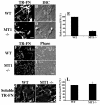
MT1-MMP is involved in endocytosis of ECM, but not soluble fibronectin. (A–I) MT1-MMP is involved in endocytosis and turnover of ECM fibronectin. MT1-MMP-null or WT cells were seeded onto pre-assembled matrix containing TR–fibronectin (A–D and F–I) or Alexa-Fluor-488–fibronectin (E) and cultured for 25–41 hours. Cells were then fixed for imaging (A–D and F–I) or processed for flow cytometry analysis (E). A and C are confocal images that were taken in a plane of focus to best show intracellular vesicles containing TR–fibronectin (arrows). Low magnification images in F and H were taken in a plane of focus to best show fibronectin fibrils. Scale bars: 20 μm (A–D) and 100 μm (F–I). (E) Flow cytometry was used to quantify endocytosed fibronectin. The data are expressed as the percentage change of MFI of endocytosed Alexa-Fluor-488–fibronectin (means ± s.e.m., n=3, P<0.0001). MFI in WT cells was set as 100%. (J–L) MT1-MMP is not required for soluble fibronectin endocytosis. MT1-MMP-null or WT cells were incubated with 10 μg/ml TR–fibronectin at 4°C for 45 minutes (pulse). Unbound fibronectin was removed and cells were then chased at 37°C for 1 hour. Endocytosed TR–fibronectin is shown in J and K (scale bar: 20 μm). Flow cytometry analysis is shown in L. The data are expressed as the percentage change of MFI of endocytosed soluble Alexa-Fluor-488–fibronectin (means ± s.e.m., n=3; P>0.05). MFI in WT cells was set as 100%.
Fig. 4.
Fig. 4.
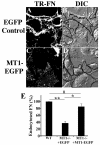
Re-expression of MT1-MMP rescues endocytosis and turnover of ECM fibronectin. MT1-MMP-null cells were seeded onto pre-assembled matrix containing TR–fibronectin (A–D) or Alexa-Fluor-633–fibronectin (E). Cells were cultured for 2 hours before being transduced with MT1-MMP–EGFP (C,D) or EGFP (A,B)-expressing adenoviruses. Cells were cultured for a total of 48 hours before fixation for confocal imaging. Scale bar: 20 μm. Cells expressing EGFP or MT1–EGFP were detected by fluorescence imaging and are outlined in B and D. Some cells were processed for flow cytometry analysis (E). The graph in E shows the percentage change of the MFI of endocytosed Alexa-Fluor-633–fibronectin. MFI in WT cells was set as 100% (means ± s.e.m., n=4; *P<0.001, **P<0.00001 and #P>0.05).
Fig. 5.
Fig. 5.
MT1-MMP is involved in endocytosis of ECM collagen I, but not ECM fibrinogen. (A–D,I–L) Pre-assembled matrix containing TR–collagen I (A–D) or Oregon-Green–fibrinogen (OG-FBN, I–L) was prepared as described in Materials and Methods. MT1-MMP-null or WT cells were cultured on pre-assembled matrices for 40–48 hours. Cells were then fixed for confocal imaging. Scale bar: 20 μm. (E–H) MT1-MMP is not required for soluble collagen I endocytosis. MT1-MMP-null or WT cells were incubated with 10 μg/ml TR–collagen I at 4°C for 50 minutes (pulse). Cells were washed and then chased at 37°C for 1 hour. Intracellular TR–collagen I was visualized by confocal imaging. Scale bar: 10 μm.
Fig. 6.
Fig. 6.
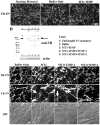
MT1-MMP enzymatic digestion rescues ECM fibronectin endocytosis and turnover. (A–C) MT1-MMP digestion does not disrupt global ECM structure. Pre-assembled matrix containing TR–fibronectin was prepared and then treated with MT1-MMP catalytic domain in the presence or absence of 50 nM TIMP-1 or TIMP-2. Matrices treated with buffer only were used as a negative control. Matrices were fixed before (A) or after (B,C) digestion. Scale bar: 40 μm. (D) Cleavage of TR-conjugated ECM fibronectin by MT1-MMP. Some matrices were collected into SDS sample buffer after digestion, and then subjected to western blot analysis using anti-Texas Red antibodies. The arrow points to full-length fibronectin. Actin was used as loading control. (E–L) MT1-MMP enzymatic digestion increases endocytosis and turnover of ECM fibronectin. MT1-MMP-null cells were cultured on MT1-MMP-enzyme-treated or control matrices for 48 hours. Cells were then fixed for confocal imaging. (E–H) TR–fibronectin (arrows point to intracellular TR–fibronectin); (E′–H′) enlarged views of the upper right sections of E–H (shown as boxed area in E). Scale bar: 20 μm. (I–L) Corresponding DIC images of E–H.
Fig. 7.
Fig. 7.
MT1-MMP regulates α5β1 integrin endocytosis. (A,B) Impaired α5β1 integrin endocytosis in MT1-MMP-null cells. Integrin endocytosis was measured using a biotinylation assay as described in the Materials and Methods. Western blot analysis using anti-α5 integrin antibodies was used to detect endocytosed α5 integrin in MT1-MMP WT (lanes 1,3,5,7) or-null (lanes 2,4,6,8) cells. Total levels of biotinlyated cell surface α5 integrin (lane 1 and 2); internalized α5 integrin after 0 minutes (lane 3 and 4) and 30 minutes (lane 5–8) of chase. In panel A, duplicate samples are shown in lanes 5 and 7; 6 and 8. (B) The relative fold change of α5 integrin endocytosis in MT1-MMP-null cells compared with that in WT cells, which was set as 1 (means ± s.e.m., n=3; P<0.0005). (C,D) Re-expression of MT1-MMP rescues α5β1 integrin endocytosis. MT1-MMP-null cells were transduced with adenoviral MT1-MMP–EGFP (lanes 3,6,9) or EGFP control (lanes 2,5,8), cultured for 48 hours, and then subjected to biotinylation assay to measure α5 integrin endocytosis. The blot was probed with anti-α5 integrin antibody. Total levels of biotinlyated cell surface α5 integrin (lane 1–3); internalized α5 integrin after 0 minutes (lane 4–6) and 30 minutes (lane 7–9) of chase. (D) The relative fold change of α5 integrin endocytosis in MT1-MMP-null cells compared with that in WT cells, which was set as 1 (means ± s.e.m., n=3, *P<0.001, #P>0.05).
Fig. 8.
Fig. 8.
Fibronectin polymerization regulates α5β1 integrin endocytosis. (A,B) Reduced α5β1 integrin endocytosis in the presence of ECM fibronectin. FN-null MFs were incubated with 10 μg/ml fibronectin in the presence of 250 nM pUR4 (FN polymerization inhibitor; lanes 1,3,5) or control peptide III-11C (lanes 2,4,6) overnight. Endocytosis of α5β1 integrin was analyzed as described in the Materials and Methods. Total levels of biotinlyated cell surface α5 integrin (lane 1 and 2); internalized α5 integrin after 0 minutes (lane 3 and 4) and 30 minutes (lane 5 and 6) of chase. (B) The relative fold change of α5 integrin endocytosis in pUR4-treated cells (+FN, +pUR4) compared with that in control cells (+FN, +III-11C), which were set as 1 (means ± s.e.m., n=3, P<0.00005). (C–E) Inhibition of fibronectin polymerization partially rescues α5β1 integrin endocytosis in MT1-MMP-null cells. MT1-MMP-null and WT cells were incubated with 250 nM pUR4 or control peptide (III-11C), and cultured for 24 hours. Endocytosis of α5β1 integrin was analyzed. (C) Western blot analysis showing total levels of biotinlyated cell surface α5 integrin (lane 1–4), and internalized α5 integrin after 0 minutes of chase (lane 5–8). (D) Levels of internalized α5 integrin after 30 minutes of chase. Duplicate samples are shown. (E) The relative fold change of α5 integrin endocytosis in cells under different treatments compared with that in WT cells treated with control peptide, which was set as 1 (means ± s.e.m., n=3; *P<0.05, **P<0.001).
Fig. 9.
Fig. 9.
Turnover of pre-existing fibronectin matrix accelerates cell migration. (A–D) FN-null MFs were cultured until 90% confluence and then supplied with 10 μg/ml fibronectin overnight. Cell monolayers were wounded, washed and then incubated with culture medium containing (+FN) or lacking (−FN) 10 μg/ml fibronectin. Representative phase images of wound areas are shown (scale bar: 20 μm). (E,F) Inhibition of fibronectin polymerization accelerates cell migration. Wound healing assay was performed as described above. Following wounding, cells were cultured in the presence or absence of 10 μg/ml fibronectin, and in the presence of 250 nM pUR4 or control peptide III-11C. The area migrated was analyzed as described in the Materials and Methods. Graphs show the relative fold change in the area migrated 4 hours (E) and 8 hours (F) post wounding by cells under various treatments to that in the presence of fibronectin, which is set to 1 (means ± s.e.m., n=3).
Fig. 10.
Fig. 10.
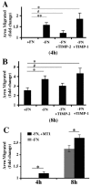
TIMP-2 blunts, whereas MT1-MMP catalytic domain further accelerates, ECM fibronectin turnover-induced cell migration. FN-null MFs were cultured until 90% confluence and then incubated with 10 μg/ml fibronectin overnight to establish a robust fibronectin matrix. Would healing assay was performed as described in the legend to Fig. 9. (A,B) Following wounding, some cells were cultured in the presence (+FN) or absence (−FN) of 10 μg/ml fibronectin, and in the presence of 50 nM TIMP-1 or TIMP-2. The graphs show the relative fold change of the area migrated by cells under various treatments to that in the presence of fibronectin, which was set as 1 (means ± s.e.m., n=3; *P<0.05, **P<0.01, #P>0.05). (A) 4 hours post wounding; (B) 8 hours post wounding. (C) After wounding, some cells were incubated with MT1-MMP catalytic domain (−FN, +MT1) or buffer control (−FN). The graph shows the relative fold change of area migrated by cells cultured in the presence of MT1-MMP to that in the absence of MT1-MMP, which was set as 1 (means ± s.e.m., n=3; *P<0.05).

Similar articles

Cited by

References

    1. Alfranca A., Lopez-Oliva J. M., Genis L., Lopez-Maderuelo D., Mirones I., Salvado D., Quesada A. J., Arroyo A. G., Redondo J. M. (2008). PGE2 induces angiogenesis via MT1-MMP-mediated activation of the TGFbeta/Alk5 signaling pathway. Blood 112, 1120-1128 - PubMed
    1. Barilla M. L., Carsons S. E. (2000). Fibronectin fragments and their role in inflammatory arthritis. Semin. Arthritis Rheum. 29, 252-265 - PubMed
    1. Berk B. C., Fujiwara K., Lehoux S. (2007). ECM remodeling in hypertensive heart disease. J. Clin. Invest 117, 568-575 - PMC - PubMed
    1. Bourdoulous S., Orend G., MacKenna D. A., Pasqualini R., Ruoslahti E. (1998). Fibronectin matrix regulates activation of RHO and CDC42 GTPases and cell cycle progression. J. Cell Biol. 143, 267-276 - PMC - PubMed
    1. Bretscher M. S. (1989). Endocytosis and recycling of the fibronectin receptor in CHO cells [published erratum appears in EMBO J. (1989). 8, 2122] EMBO. J. 8, 1341-1348 - PMC - PubMed

Publication types

MeSH terms