. 2011 Dec 1;124(Pt 23):3956-66.
doi: 10.1242/jcs.080374. Epub 2011 Dec 8.

Cell organization, growth, and neural and cardiac development require αII-spectrin

Affiliations

Cell organization, growth, and neural and cardiac development require αII-spectrin

Michael C Stankewich et al. J Cell Sci. .

Abstract

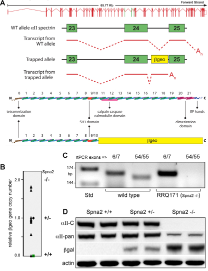
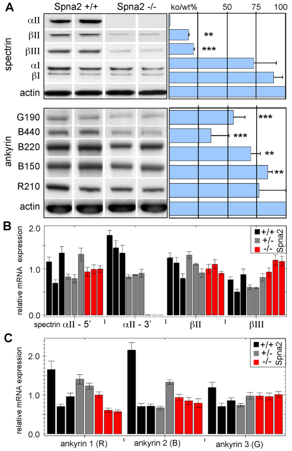
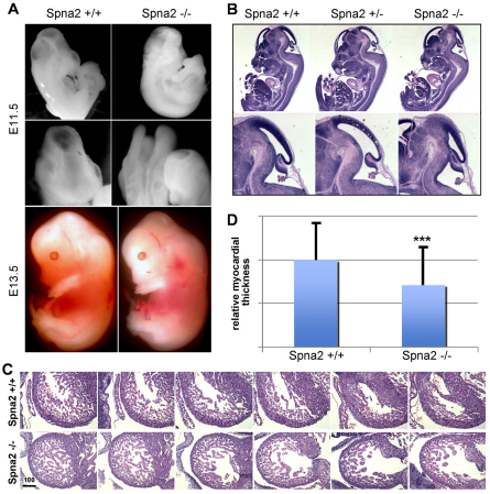
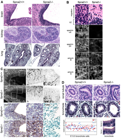
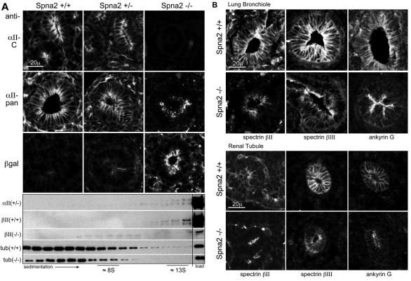
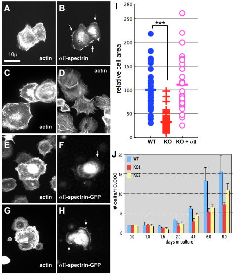
Spectrin α2 (αII-spectrin) is a scaffolding protein encoded by the Spna2 gene and constitutively expressed in most tissues. Exon trapping of Spna2 in C57BL/6 mice allowed targeted disruption of αII-spectrin. Heterozygous animals displayed no phenotype by 2 years of age. Homozygous deletion of Spna2 was embryonic lethal at embryonic day 12.5 to 16.5 with retarded intrauterine growth, and craniofacial, neural tube and cardiac anomalies. The loss of αII-spectrin did not alter the levels of αI- or βI-spectrin, or the transcriptional levels of any β-spectrin or any ankyrin, but secondarily reduced by about 80% the steady state protein levels of βII- and βIII-spectrin. Residual βII- and βIII-spectrin and ankyrins B and G were concentrated at the apical membrane of bronchial and renal epithelial cells, without impacting cell morphology. Neuroepithelial cells in the developing brain were more concentrated and more proliferative in the ventricular zone than normal; axon formation was also impaired. Embryonic fibroblasts cultured on fibronectin from E14.5 (Spna2(-/-)) animals displayed impaired growth and spreading, a spiky morphology, and sparse lamellipodia without cortical actin. These data indicate that the spectrin-ankyrin scaffold is crucial in vertebrates for cell spreading, tissue patterning and organ development, particularly in the developing brain and heart, but is not required for cell viability.


Figures

Fig. 1.
Fig. 1.
Targeted disruption of αII-spectrin. (A) An ES cell line was established using gene trap vector RRQ171 (β-geo) from Bay Genomics. Analysis by 5′ RACE identified insertion of the vector between exons 24 and 25 of the murine Spna2 gene. This created a fusion transcript with a spectrin message truncated by the addition of β-geo. A cartoon of this fusion transcript, and the anticipated fusion protein, is depicted. (B) E11.5 embryos derived from a RRQ171 heterozygous breeding pairs were genotyped by quantitative RT-PCR for β-geo. (C) RT-PCR analysis with intron-bridging primer pairs directed to upstream exons (6/7) or downstream exons (54/55) confirmed the absence of mRNA encoding full-length αII-spectrin in homozygotes. (D) Western blot analysis showed that monoclonal antibodies to αII-spectrin (αII-C) that react with peptide sequences downstream of the exon trap were negative in homozygotes. Pan-reactive anti-spectrin antibodies (αII-pan) detect the fusion protein. Antibodies to β-gal confirm the presence of the fusion protein in both the homo- and heterozygotes.
Fig. 2.
Fig. 2.
Loss of αII-spectrin destabilizes βII- and βIII-spectrin. (A) Western blot analysis of whole embryos comparing the relative steady state protein levels of several spectrins and ankyrins. Each lane represents results with a separate embryo. The loss of αII-spectrin reduced the steady state levels of βII- and βIII-spectrin to below 20% of normal; αI- and βI-spectrin were unchanged. The abundance of βIV- and βV-spectrin was too low in embryos younger than E14.5 to be reliably evaluated. Ankyrins B440 and G190 were both significantly diminished, as were ankyrins B220 and B150, albeit not to the same degree as the β-spectrins. (B,C) Quantitative RT-PCR analysis revealed that despite the change in protein levels, there were no consistent changes in the mRNA levels of any spectrin or ankyrin (except for the disrupted Spna2 gene, as measured with primers targeted to the 3′ end, αII-3′). Each analysis was performed in triplicate on two or three separate animals. Error bars show ±1 s.d. **P<0.05, ***P<0.005.
Fig. 3.
Fig. 3.
Spna2−/− embryos die at E12.5–16.5 with multiple defects. (A,B) Whole mounts depicting the gross morphological defects in head development, with frequent neural tube closure defects (E11.5 embryos shown). In cases where the neural tube closed, there remained distinct alterations in head and back curvature with craniofacial abnormalities. (C) Serial sections through the fetal heart. The Spna2−/− hearts have an irregular shaped ventricle, and a thinned compact myocardium. Scale bar: 100 μm. (D) Quantitative comparison of cardiac wall thickness. On average, the myocardium of the Spna2−/− hearts was 70.9% of the thickness of normal hearts of the same gestational age. This difference is highly significant (n=101; ***P=4.2×10⁻¹³). Error bars indicate s.d.
Fig. 4.
Fig. 4.
Histology of Spna2−/− embryos. (A) The VZ and SVZ of the developing brain display significant histological changes. Wild-type embryos at this stage of development display a thick pseudostratified VZ with active upward migration and population of overlying layers from the SVZ. In the Spna2−/− animals, the VZ was more compact, with fewer cells filling the more superficial layers (boxed area, shown enlarged in B). Other organs revealed no significant histological changes; kidney and lung are shown. In these organs, tubules and bronchioles were developing normally. (B) Enlargement of area outlined in A. In wild-type animals, αII- and βII-spectrin are rich in axon bundles (boxed) and are also present at synaptic terminals and initial axon segments (arrows). Ankyrin B is associated with unmyelinated axonal processes in the embryonic mouse (Chan et al., 1993), whereas ankyrin G is found along axons and in dendrites and concentrates in the initial axon segments (Kordeli et al., 1995). The loss of αII-spectrin disrupts the localization of these proteins, especially along the axon bundles, which no longer are marked by either spectrin or ankyrin. (C) Top two rows: Staining of sections with anti-tau demonstrates developing axon bundles in the VZ. Magnified images (3×) of the boxed areas are shown on the right with inverse contrast. Note the foreshortened, sparse and more disordered axon segments and increased concentration of tau near the soma when αII-spectrin is disrupted. Bottom two rows: Immunostaining for Ki67, a proliferation marker, reveals increased proliferation with spectrin loss. Averaged over multiple fields, and scoring any detected Ki67 staining as positive, 34±11% of cells in the wild-type animals were positive as compared with 46±8% of the cells in the spectrin-deleted animals. This represents a highly significant (P<0.006) 35% increase in proliferative activity. TUNEL staining (right) reveals no change in the level of apoptosis. (D) Higher power views of developing renal tubules and lung bronchioles indicate preservation of epithelial morphology. Comparison of epithelial cell height reveals no changes, even without laterally associated spectrin or ankyrin (see Fig. 5).
Fig. 5.
Fig. 5.
Loss of αII-spectrin leads to a redistribution of both βII- and βIII-spectrin and ankyrin. (A) In developing renal tubules, αII-spectrin is distributed uniformly over both the basolateral and apical surfaces in wild-type (Spna2+/+) embryos, as detected by both the C-terminal anti-αII-spectrin antibody (αII-C) as well as the pan-reactive spectrin antibody (αII-pan). In the Spna2−/− tubules, the residual spectrin–β-gal fusion protein is totally lost from the basolateral membrane, and concentrated in coarse apical and sub-apical pools as detected by both αII-pan and by an antibody to β-gal. In heterozygotes (Spna2+/−), the presence of the fusion protein had no detectable effect on the distribution of wild-type αII-spectrin, nor was the distribution of the fusion protein changed by the presence of αII-spectrin. Below: Sucrose density gradient analysis of proteins extracted from E13.5 embryos. In the homozygote (Spna2−/−), βII-spectrin does not sediment with the high molecular weight protein complex at 13S. (B) Immunofluorescent micrographs of bronchioles and renal tubules. Without intact αII-spectrin, βII- and βIII-spectrin and ankyrin G are largely absent from the lateral membrane of both renal and bronchiole epithelial cells. Residual βII- and βIII-spectrin and ankyrin G concentrate instead at the apical membrane in a pattern similar to that of the αII-spectrin–β-gal fusion protein.
Fig. 6.
Fig. 6.
MEFs lacking intact αII-spectrin display impaired growth and spreading, and altered morphology. (A,B) MEFs from E14.5 embryos were plated onto fibronectin, grown for up to 3 hours, and stained as indicated. The images shown here were from cells examined at 30 minutes after replating. (A) MEF from wild-type cells, stained for F-actin with rhodamine phalloidin. Note the peripheral cortical and perinuclear actin, cell spreading, and abundant ruffling edges and lamellipodia. (B) Same wild-type cells as in A, stained for αII-spectrin. Note the coincidence of spectrin with the ruffling edge (arrows). (C,D) MEFs from Spna2+/+ (C) or Spna2−/− (D) embryos under same conditions as in A, stained for actin. Note the lack of spreading and lamellipodia, and abundant filopodial projections and stress filaments in the spectrin-deficient cells. (EH) MEFs from Spna2−/− embryos under same conditions as A, but transfected with wild-type GFP-labeled αII-spectrin. Double immunofluorescent images are shown, stained for actin or GFP. Arrows mark the accumulation of GFP–spectrin at the ruffling edge and lamellipodia. Note the recovery of cortical and perinuclear actin, ruffling, and spreading in the transfected cells expressing GFP–αII-spectrin. (I) Scatter diagram of individual cell areas of MEFs from Spna2+/+ (WT) or Spna2−/− (KO) embryos, or from Spna2−/− cells transfected with GFP–αII-spectrin (KO+αII). The KO cells are significantly less spread, with an average area just 32% of wild-type cells (***P≤1.6×10⁻¹⁶); there is no difference in area between the wild-type cells and the Spna2−/− cells transfected with GFP–αII-spectrin. (J) Comparison of cell growth in culture, as measured by cell count, of wild-type and two MEF lines (KO1, KO2) independently derived from separate Spna2−/− embryos.

Similar articles

Cited by

References

    1. Bellenchi G. C., Gurniak C. B., Perlas E., Middei S., Ammassari-Teule M., Witke W. (2007). N-cofilin is associated with neuronal migration disorders and cell cycle control in the cerebral cortex. Genes. Dev. 21, 2347-2357 - PMC - PubMed
    1. Bennett V., Baines A. J. (2001). Spectrin and ankyrin-based pathways: metazoan inventions for integrating cells into tissues. Physiol. Rev. 81, 1353-1392 - PubMed
    1. Bennett V., Healy J. (2008). Organizing the fluid membrane bilayer: diseases linked to spectrin and ankyrin. Trends Mol. Med. 14, 28-36 - PubMed
    1. Bialkowska K., Saido T. C., Fox J. E. (2005). SH3 domain of spectrin participates in the activation of Rac in specialized calpain-induced integrin signaling complexes. J. Cell Sci. 118, 381-395 - PubMed
    1. Bloch R. J., Morrow J. S. (1989). An unusual β-spectrin associated with clustered acetylcholine receptors. J. Cell Biol. 108, 481-493 - PMC - PubMed

Publication types

MeSH terms