. 2011 Dec 1;124(Pt 23):3967-79.
doi: 10.1242/jcs.081372. Epub 2011 Dec 8.

WRN helicase regulates the ATR-CHK1-induced S-phase checkpoint pathway in response to topoisomerase-I-DNA covalent complexes

Affiliations

WRN helicase regulates the ATR-CHK1-induced S-phase checkpoint pathway in response to topoisomerase-I-DNA covalent complexes

Birija Sankar Patro et al. J Cell Sci. .

Abstract

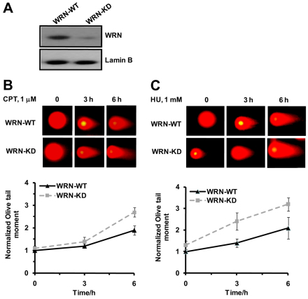
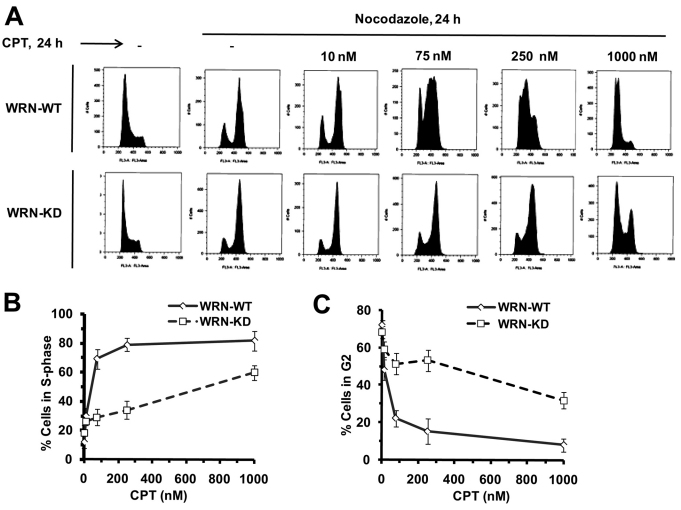
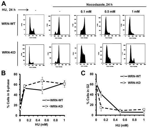
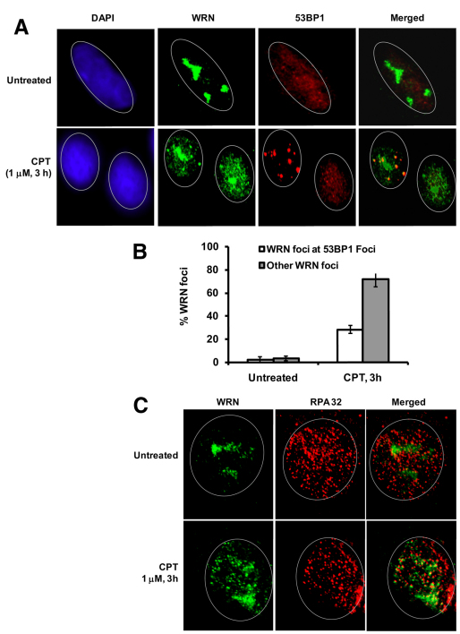
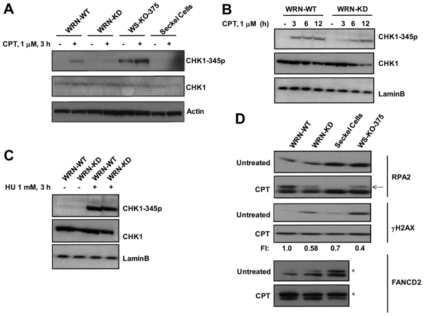
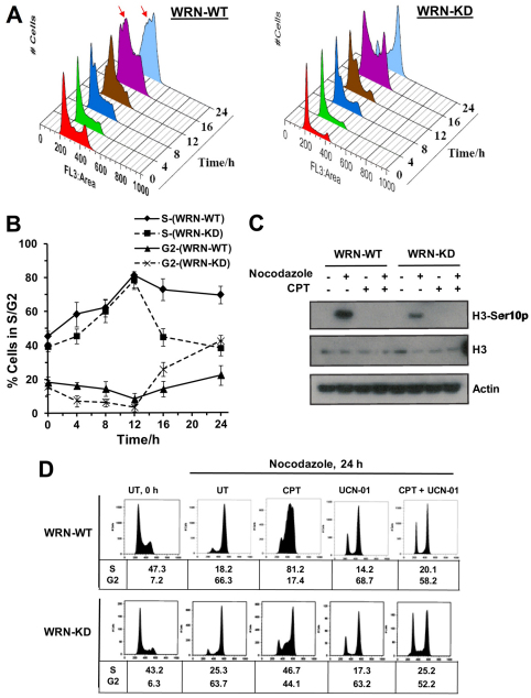
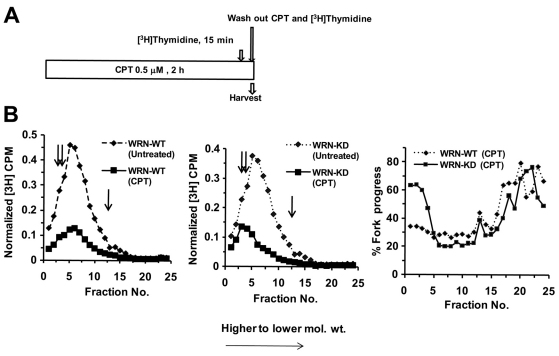
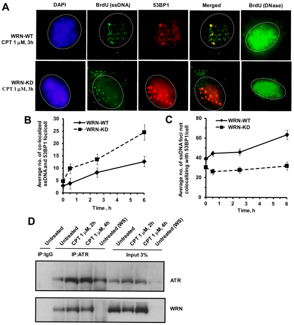
Checkpoints are cellular surveillance and signaling pathways that coordinate the response to DNA damage and replicative stress. Consequently, failure of cellular checkpoints increases susceptibility to DNA damage and can lead to profound genome instability. This study examines the role of a human RECQ helicase, WRN, in checkpoint activation in response to DNA damage. Mutations in WRN lead to genomic instability and the premature aging condition Werner syndrome. Here, the role of WRN in a DNA-damage-induced checkpoint was analyzed in U-2 OS (WRN wild type) and isogenic cells stably expressing WRN-targeted shRNA (WRN knockdown). The results of our studies suggest that WRN has a crucial role in inducing an S-phase checkpoint in cells exposed to the topoisomerase I inhibitor campthothecin (CPT), but not in cells exposed to hydroxyurea. Intriguingly, WRN decreases the rate of replication fork elongation, increases the accumulation of ssDNA and stimulates phosphorylation of CHK1, which releases CHK1 from chromatin in CPT-treated cells. Importantly, knockdown of WRN expression abolished or delayed all these processes in response to CPT. Together, our results strongly suggest an essential regulatory role for WRN in controlling the ATR-CHK1-mediated S-phase checkpoint in CPT-treated cells.


Figures

Fig. 1.
Fig. 1.
Depletion of WRN in U-2 OS cells leads to increased accumulation of DSBs in response to DNA-damaging agents. (A) Western blot analysis shows ~80% reduction in the level of WRN protein expression in WRN-KD cells compared with WRN-WT cells. Lamin B was used as a protein loading control. Analysis of DNA DSBs in WRN-WT and WRN-KD cells in response to (B) CPT and (C) HU, respectively. Cells were either left untreated or treated with CPT (1 μM) and HU (1 mM) and DNA DSBs were evaluated by neutral comet assay after 3 hours and 6 hours of respective drug treatment. Lower panels in B and C show the quantification of DSBs in terms of Olive tail moment in comet assay. Points represent mean ± s.d. from at least three experiments (200 nuclei in each experiment).
Fig. 2.
Fig. 2.
Depletion of WRN causes defective S-phase checkpoint in the presence of CPT. (A) WRN-WT and WRN-KD cells were treated with increasing concentration of CPT (0–1000 nM) for 24 hours in the presence of nocodazole (0.25 μg/ml) as indicated. After 24 hours cells were harvested to determine cell cycle distributions by flow cytometry as shown in cell count vs DNA content profiles of the cells. (B,C) Quantification of S-phase population (B) and G2–M-phase population (C) in the same experiments as described in Fig. 1A (n=4). Points represent mean ± s.d.
Fig. 3.
Fig. 3.
WRN is dispensable for HU-induced checkpoint. (A) WRN-WT and WRN-KD cells were treated with increasing concentrations of HU (0.1–1 mM) for 24 hours in the presence of nocodazole (0.25 μg/ml) as indicated. After 24 hours, cells were harvested to determine cell cycle distributions by flow cytometry as shown in cell count vs DNA content profiles of the cells. (B,C) Quantification of S-phase population (B) and G2–M-phase population (C) in the same experiments as described in Fig. 3A (n=3). Points represent mean ± s.d.
Fig. 4.
Fig. 4.
DSB-dependent and -independent localization of WRN in response to CPT. (A) WRN and 53BP1 colocalize to nuclear foci. Indirect immunofluorescence of WRN (green) and 53BP1 (red) in untreated and CPT (1 μM, 3 hours) treated cells are shown. In the merged image, yellow coloration is due to colocalization of WRN and 53BP1. (B) Quantification of WRN foci formation at 53BP1 sites (DSBs) and other sites (non-DSB) in the same experiment as described in A. Bars represent mean ± s.d. (C) Detection of WRN foci formation at replication sites by immunostaining. Indirect immunofluorescence of WRN (green) and RPA32 (red) in untreated and CPT treated cells (1 μM, 3 hours) are shown. In the merged image, yellow coloration is due to colocalization of WRN and RPA32. The DAPI staining (blue) in left panel shows the position of the nucleus. A minimum of 150 cells were assessed for each sample in three independent experiments.
Fig. 5.
Fig. 5.
WRN regulates ATR-mediated signaling in response to CPT. (A) CHK1 phosphorylation on Ser345 was detected in WRN-WT, WRN-KD, WS-KO-375 cells and Seckel cells treated with CPT (1 μM). Cells were harvested at the indicated time point and cell lysates prepared as described for western analyses. (B) CHK1 phosphorylation was determined at various time points as indicated. (C) CHK1 phosphorylation on Ser345 was detected in control and WRN-KD cells treated with HU (1 mM, 3 hours). (D) Phosphorylation and ubiquitylation of ATR substrates in control and WRN-KD cells after CPT treatment (1 μM, 3 hours). Arrow shows phosphorylated form of RPA32; asterisk indicates monoubiquitylated form of FANCD2. Because endogenous levels of γH2AX and monoubiquitylated FANCD2 are high in WRN-depleted cells at untreated conditions, fold increase (FI) in the levels of induced γH2AX and monoubiquitylated FANCD2 in response to CPT are given by normalizing the fold increase (FI) in WRN-WT cells to 1.
Fig. 6.
Fig. 6.
WRN regulates CPT-induced intra-S-phase checkpoint but not G2 checkpoint. (A) WRN-WT and WRN-KD cells were treated with CPT (0.25 μM) and cells were harvested at indicated time points to determine cell cycle distributions by flow cytometry. Arrows indicate arrested cells in S phase. (B) Quantification of S-phase and G2–M-phase populations in A (n=3). Points represent mean ± s.d. (C) Histone H3 Ser10 phosphorylation was quantified in WRN-WT and WRN-KD cells untreated and treated with CPT and nocodazole alone and together as indicated. Cell lysates were prepared at the indicated time point. (D) WRN-WT and WRN-KD cells were untreated (UT) or treated with CPT (0.25 μM) and nocodazole (0.25 μg/ml) for 24 hours in the presence or absence of UCN 01 (0.1 μM) as indicated. Cell cycle distribution was analyzed by flow cytometry as shown in cell count vs DNA content profiles of the cells. Percentage of cells in S and G2 phase population is indicated under respective samples (n=3).
Fig. 7.
Fig. 7.
Inhibition of chain elongation of DNA replication is impaired in WRN-depleted cells following exposure to CPT. (A) Schematic representation of [3H]thymidine and CPT treatment. (B) Cells were harvested after CPT treatment as in A, and nascent DNA was separated by velocity sedimentation. Net 3H radioactivity corrected for 14C spillover was normalized to cell number (total 14C radioactivity). Sedimentation is from right to left (i.e. fraction 1 is at the bottom of the gradient and fraction 25 at the top). Percentage fork progress in the last panel was calculated by dividing the value of normalized 3H c.p.m. of each CPT-treated fraction with that of untreated sample and multiplied by 100. The double and single arrows indicate the sedimentation rates of 167 kb and 37 kb DNA marker, respectively.
Fig. 8.
Fig. 8.
WRN is involved in the generation of ssDNA and ATR activation in response to CPT. (A) Detection of incorporated BrdU specifically in ssDNA after CPT treatment. Cells pre-labeled with BrdU were treated with CPT (1 μM, 3 hours) and stained with antibodies against BrdU (green) and 53BP1 (red). The DAPI staining (blue) in the left panel shows the position of the nucleus. DNase-treated cells are included to show that the whole-cell population was uniformly labeled with BrdU (Syljuasen et al., 2005; Lee et al., 2006). (B,C) Quantification of ssDNA foci formation at DSBs and non-DSB sites in the same experiment as described in A at indicated time points. A minimum of 100–200 cells were assessed for each sample in three independent experiments. Points represent mean ± s.d. (D) WRN co-immunoprecipitated with ATR in human U-2 OS cells. Immunoprecipitations were carried out using anti-ATR antibody, or an immunoglobulin G control in presence of ethidium bromide (30 μg/ml), on extracts from cells exposed to no treatment or CPT treatment (1 μM, 2 and 4 hours). The immunoprecipitates were analyzed by western blot for the presence of WRN using anti-WRN antibodies. WS, WS-KO-375 cells.

Similar articles

Cited by

References

    1. Ahn B., Lee J. W., Jung H., Beck G., Bohr V. A. (2009). Mechanism of Werner DNA helicase: POT1 and RPA Stimulates WRN to unwind beyond gaps in the translocating Strand. PLoS One 4, e4673 - PMC - PubMed
    1. Ammazzalorso F., Pirzio L. M., Bignami M., Franchitto A., Pichierri P. (2010). ATR and ATM differently regulate WRN to prevent DSBs at stalled replication forks and promote replication fork recovery. EMBO J. 29, 3156-3169 - PMC - PubMed
    1. Andreassen P. R., D'Andrea A. D., Taniguchi T. (2004). ATR couples FANCD2 monoubiquitination to the DNA-damage response. Genes Dev. 18, 1958-1963 - PMC - PubMed
    1. Baynton K., Otterlei M., Bjørås M., Kobbe C., Bohr V. A., Seeberg E. (2003). WRN interacts physically and functionally with the recombination mediator protein RAD52. J. Biol. Chem. 278, 36476-36486 - PubMed
    1. Bernstein K. A., Shor E., Sunjevaric I., Fumasoni M., Burgess R. C., Foiani M., Branzei D., Rothstein R. (2009). Sgs1 function in the repair of DNA replication intermediates is separable from its role in homologous recombinational repair. EMBO J. 28, 915-925 - PMC - PubMed

Publication types

MeSH terms