. 2012 Jul;61(7):1065-80.
doi: 10.1007/s00262-011-1165-5. Epub 2011 Dec 13.

Activation-induced cell death of memory CD8+ T cells from pleural effusion of lung cancer patients is mediated by the type II Fas-induced apoptotic pathway

Affiliations

Activation-induced cell death of memory CD8+ T cells from pleural effusion of lung cancer patients is mediated by the type II Fas-induced apoptotic pathway

Heriberto Prado-Garcia et al. Cancer Immunol Immunother. 2012 Jul.

Abstract

Lung cancer is the second most common form of cancer and the leading cause of cancer death worldwide. Pleural effusions, containing high numbers of mononuclear and tumor cells, are frequent in patients with advanced stages of lung cancer. We reported that in pleural effusions from primary lung cancer, the CD8+ T cell subpopulation, and particularly the terminally differentiated subset, is reduced compared to that of non-malignant effusions. We analyzed the participation of activation-induced cell death (AICD) and extrinsic pathways (type I or II) as mechanisms for the decrease in pleural effusion CD8+ T cell subpopulation. Pleural effusion or peripheral blood CD4+ and CD8+ T cells, from lung cancer patients, were stimulated with anti-CD3 antibody and analyzed for (a) apoptosis by annexin-V-binding and TUNEL assay, (b) transcript levels of Fas ligand (FasL) and TRAIL by real-time RT-PCR, (c) expression of FasL and TRAIL, measured as integrated mean fluorescence intensities (iMFI) by flow cytometry, (d) expression of Bcl-2 and BIM molecules, measured as MFI, and (e) apoptosis inhibition using caspase-8 and -9 inhibitors. Pleural effusion CD8+ T cells, but not CD4+ T cells, from cancer patients underwent AICD. Blocking FasL/Fas pathway protected from AICD. Upregulation of FasL and TRAIL expressions was found in pleural effusion CD8+ T cells, which also showed a subset of Bcl-2 low cells. In memory CD8+ T cells, AICD depended on both extrinsic and intrinsic apoptotic pathways. Hence, in the pleural space of lung cancer patients, AICD might compromise the antitumor function of CD8+ T cells.


Figures

Fig. 1
Fig. 1
CD4+ and CD8+ T cells from lung cancer patients do not undergo apoptosis after treatment with agonistic anti-Fas mAb. a Percentages of spontaneous apoptosis in CD4+ and CD8+ T cells from lung cancer and control groups after 24-h incubation of PEMCs or PBMCs in RPMI-1640 medium, determined by annexin-V binding. Bars depict the means. b PEMCs or PBMCs were incubated with anti-Fas mAb (1 μg/ml, clone IgM, clone CH11) or isotype control for 24 h; then, percentages of apoptotic cells were determined by annexin-V binding in CD4+ and CD8+ T cells from pleural effusion (PE) and peripheral blood (PB) of lung cancer patients (n = 12)
Fig. 2
Fig. 2
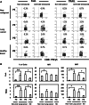
Pleural effusion CD8+ T cells from lung cancer patients stimulated with anti-CD3 mAb presented high levels of apoptosis. a Plots showing the gating strategies for lymphocytes, cells were gated on the corresponding CD4+ or CD8+ T cell subpopulation, and analysis of annexin-V versus propidium iodide (PI) was performed, data from CD3-stimulated and non-stimulated pleural effusion (PE) and peripheral blood (PB) CD4+ and CD8+ T cells from a cancer patient. b Comparison of apoptosis induced by anti-CD3 mAb stimulation (10 μg/ml) in CD4+ and CD8+ T cells from all groups. PE from non-malignant patients (n = 11), PE and peripheral blood (PB) from lung cancer patients (n = 15) and PB from healthy donors (n = 13). Bars depict the mean ± SE
Fig. 3
Fig. 3
AICD of pleural effusion CD8+ T cells from lung cancer patients is blocked by caspase-3 inhibitor or FasL mAb. a Data from pleural effusion CD4+ and CD8+ T cells from a cancer patient are shown. PEMCs were stimulated with anti-CD3 mAbs for 30 h in the presence of a caspase-3 inhibitor, blocking anti-FasL mAb or isotype control. TUNEL-positive cells were determined by TUNEL versus FSC 5% contour outlier plot of CD4+ (lower row) or CD8+ cells (upper row), gated from CD3+ cell population, as described in “Materials and methods”. Percentage of TUNEL-positive cells is shown. b Comparison of apoptosis induced by anti-CD3 mAb stimulation (10 μg/ml) in pleural effusion CD8+ T cells. Percentage of anti-CD3-induced apoptosis is shown for isotype control, caspase-3 inhibitor (C-3 inh), and anti-FasL mAb (n = 5). Bars depict the mean ± SE
Fig. 4
Fig. 4
The expression of FasL and TRAIL is increased in anti-CD3-stimulated CD8+ T cells from lung cancer patients compared to non-malignant controls. a Representative 5% contour outlier plots showing the percentages of FasL- or TRAIL-positive CD8+ T cells from PEMC or PBMC of a cancer patient and controls under non-stimulated and anti-CD3-stimulated conditions. b Comparison of FasL (upper row) and TRAIL (lower row) production, the percentage of cells (left), MFI (middle), and iMFI (right) of FasL or TRAIL expressing CD8+ T cells induced by anti-CD3 mAb stimulation (10 μg/ml) from all groups is shown. PE from non-malignant patients (Non-mal, n = 11), PE and PB from lung cancer patients (n = 15) and PB from healthy donors (HD, n = 13). Bars depict the mean ± SE, *P < 0.0001, **P < 0.001, + P < 0.01
Fig. 5
Fig. 5
Pleural effusion CD8+ T cells from lung cancer patients stimulated with anti-CD3 mAbs upregulate FasL and TRAIL mRNA levels. PE and PB CD8+ T cells from lung cancer patients and PB CD8+ T cells from healthy subjects (HD) were purified by negative selection using magnetic beads. Cell stimulation was performed by incubating with anti-CD3 mAb for 4 h. Total RNA was extracted, and qRT–PCR performed. The results were analyzed by the comparative Ct method, using β-actin as an internal control (see “Materials and methods”). The results are semiquantitative and represent the n-fold difference of the transcript levels in a particular sample compared to calibrator cDNA (cDNA samples from non-stimulated CD8+ T cells from each subject). Bars depict the mean
Fig. 6
Fig. 6
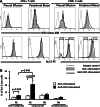
A Bcl-2lo subset is induced in pleural effusion CD8+, but not CD4+, T cells from lung cancer patients upon anti-CD3-stimulation. a BIM and Bcl-2 expression levels were analyzed in cells stimulated with anti-CD3 mAb (black line) and non-stimulated (gray area) for 24 h. Histograms obtained from CD4+ and CD8+ T cells from a lung cancer patient are shown. Isotype control is indicated by a gray line. b Percentages of Bcl-2lo cells in CD8+ T cells from lung cancer and control groups determined by analysis of Bcl-2 low region (n = 7). Bars depict the mean ± SE
Fig. 7
Fig. 7
CD8+ T cell subsets from malignant effusions are susceptible to apoptosis after CD3 stimulation. a Distribution of memory, terminally differentiated and naïve CD8+ T cell subsets and corresponding annexin-V histograms under non-stimulated and stimulated conditions. Percentages of annexin-V-positive cells are shown. b Percentages of annexin-V-positive cells in pleural effusion CD8+ T cell subsets from lung cancer patients (n = 7). Bars depict the mean ± SE
Fig. 8
Fig. 8
Caspase-8 and -9 inhibitors rescued memory CD8+ T cells from AICD. a Data from pleural effusion CD8+ T cells from a cancer patient are shown. PEMCs were stimulated with anti-CD3 mAbs in the presence of a caspase-8 inhibitor, a caspase-9 inhibitor, or both. Annexin-V-positive cells were analyzed by histograms within the PI CD8+ T cell population as described in “Materials and methods”. Percentages of anti-CD3-induced apoptosis are shown for each condition (n = 9). b Representative analysis of memory CD8+ T cells. PEMCs were stimulated with anti-CD3 mAbs in the presence of a caspase-8 inhibitor, a caspase-9 inhibitor, or both. Percentages of annexin-V-positive cells were then determined in memory, naïve, and terminally differentiated subsets. Percentages of anti-CD3-induced apoptosis are shown for each condition (n = 5). *P < 0.05 with respect to cells without inhibitor (w/o inh), + P < 0.001 with respect to cells without inhibitor (w/o inh)

Similar articles

Cited by

References

    1. Alberg AJ, Ford JG, Samet JM. Epidemiology of lung cancer: ACCP evidence-based clinical practice guidelines (2nd edition) Chest. 2007;132:29S–55S. doi: 10.1378/chest.07-1347. - DOI - PubMed
    1. Jemal A, Siegel R, Xu J, Ward E. Cancer statistics. CA Cancer J Clin. 2008;58:71–96. doi: 10.3322/CA.2007.0010. - DOI - PubMed
    1. Atanackovic D, Block A, de Weerth A, Faltz C, Hossfeld DK, Hegewisch-Becker S. Characterization of effusion-infiltrating T cells: benign versus malignant effusions. Clin Cancer Res. 2004;10:2600–2608. doi: 10.1158/1078-0432.CCR-03-0239. - DOI - PubMed
    1. Pace E, Bruno TF, Berenger B, Mody CH, Melis M, Ferraro M, Tipa A, Bruno A, Profita M, Bonsignore G, Gjomarkaj M. Elevated expression of prostaglandin receptor and increased release of prostaglandin E2 maintain the survival of CD45RO+ T cells in the inflamed human pleural space. Immunology. 2007;121:427–436. doi: 10.1111/j.1365-2567.2007.02593.x. - DOI - PMC - PubMed
    1. Prado-Garcia H, Aguilar-Cazares D, Flores-Vergara H, Mandoki JJ, Lopez-Gonzalez JS. Effector, memory and naïve CD8+ T cells in peripheral blood and pleural effusion from lung adenocarcinoma patients. Lung Cancer. 2005;47:361–371. doi: 10.1016/j.lungcan.2004.07.046. - DOI - PubMed

Publication types

Substances