

Profilin 1 is a potential biomarker for bladder cancer aggressiveness
- PMID: 22159600
- PMCID: PMC3322560
- DOI: 10.1074/mcp.M111.009449
Profilin 1 is a potential biomarker for bladder cancer aggressiveness
Abstract
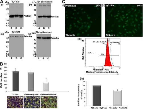
Of the most important clinical needs for bladder cancer (BC) management is the identification of biomarkers for disease aggressiveness. Urine is a "gold mine" for biomarker discovery, nevertheless, with multiple proteins being in low amounts, urine proteomics becomes challenging. In the present study we applied a fractionation strategy of urinary proteins based on the use of immobilized metal affinity chromatography for the discovery of biomarkers for aggressive BC. Urine samples from patients with non invasive (two pools) and invasive (two pools) BC were subjected to immobilized metal affinity chromatography fractionation and eluted proteins analyzed by 1D-SDS-PAGE, band excision and liquid chromatography tandem MS. Among the identified proteins, multiple corresponded to proteins with affinity for metals and/or reported to be phosphorylated and included proteins with demonstrated association with BC such as MMP9, fibrinogen forms, and clusterin. In agreement to the immobilized metal affinity chromatography results, aminopeptidase N, profilin 1, and myeloblastin were further found to be differentially expressed in urine from patients with invasive compared with non invasive BC and benign controls, by Western blot or Elisa analysis, nevertheless exhibiting high interindividual variability. By tissue microarray analysis, profilin 1 was found to have a marked decrease of expression in the epithelial cells of the invasive (T2+) versus high risk non invasive (T1G3) tumors with occasional expression in stroma; importantly, this pattern strongly correlated with poor prognosis and increased mortality. The functional relevance of profilin 1 was investigated in the T24 BC cells where blockage of the protein by the use of antibodies resulted in decreased cell motility with concomitant decrease in actin polymerization. Collectively, our study involves the application of a fractionation method of urinary proteins and as one main result of this analysis reveals the association of profilin 1 with BC paving the way for its further investigation in BC stratification.
Figures


Similar articles
-
IMAC fractionation in combination with LC-MS reveals H2B and NIF-1 peptides as potential bladder cancer biomarkers.J Proteome Res. 2013 Sep 6;12(9):3969-79. doi: 10.1021/pr400255h. Epub 2013 Aug 21. J Proteome Res. 2013. PMID: 23924207
-
LC-MS metabolomics of urine reveals distinct profiles for non-muscle-invasive and muscle-invasive bladder cancer.World J Urol. 2022 Oct;40(10):2387-2398. doi: 10.1007/s00345-022-04136-7. Epub 2022 Sep 4. World J Urol. 2022. PMID: 36057894 Review.
-
Comparative Tissue Proteomics of Microdissected Specimens Reveals Novel Candidate Biomarkers of Bladder Cancer.Mol Cell Proteomics. 2015 Sep;14(9):2466-78. doi: 10.1074/mcp.M115.051524. Epub 2015 Jun 16. Mol Cell Proteomics. 2015. PMID: 26081836 Free PMC article.
-
Silencing of Profilin-1 suppresses cell adhesion and tumor growth via predicted alterations in integrin and Ca2+ signaling in T24M-based bladder cancer models.Oncotarget. 2016 Oct 25;7(43):70750-70768. doi: 10.18632/oncotarget.12218. Oncotarget. 2016. PMID: 27683119 Free PMC article.
-
Urinary markers in the everyday diagnosis of bladder cancer.Urologia. 2013 Sep-Dec;80(4):265-75. doi: 10.5301/urologia.5000041. Epub 2013 Nov 29. Urologia. 2013. PMID: 24419920 Review.
Cited by
-
A simple urine test by 3D-plus-3D immunoassay guides precise in vitro cancer diagnosis.Bioeng Transl Med. 2023 Jan 18;8(3):e10489. doi: 10.1002/btm2.10489. eCollection 2023 May. Bioeng Transl Med. 2023. PMID: 37206218 Free PMC article.
-
Proteomic studies of urinary biomarkers for prostate, bladder and kidney cancers.Nat Rev Urol. 2013 Apr;10(4):206-18. doi: 10.1038/nrurol.2013.24. Epub 2013 Feb 26. Nat Rev Urol. 2013. PMID: 23443013 Review.
-
Profilin: many facets of a small protein.Biophys Rev. 2020 Aug;12(4):827-849. doi: 10.1007/s12551-020-00723-3. Epub 2020 Jul 13. Biophys Rev. 2020. PMID: 32661903 Free PMC article. Review.
-
Contextualised urinary biomarker analysis facilitates diagnosis of paediatric obstructive sleep apnoea.Sleep Med. 2014 May;15(5):541-9. doi: 10.1016/j.sleep.2014.01.010. Epub 2014 Feb 7. Sleep Med. 2014. PMID: 24726570 Free PMC article.
-
Recent progress in mass spectrometry-based urinary proteomics.Clin Proteomics. 2024 Feb 22;21(1):14. doi: 10.1186/s12014-024-09462-z. Clin Proteomics. 2024. PMID: 38389064 Free PMC article. Review.
References
-
- Dinney C. P., McConkey D. J., Millikan R. E., Wu X., Bar-Eli M., Adam L., Kamat A. M., Siefker-Radtke A. O., Tuziak T., Sabichi A. L., Grossman H. B., Benedict W. F., Czerniak B. (2004) Focus on bladder cancer. Cancer Cell 6, 111–116 - PubMed
-
- Parkin D. M. (2008) The global burden of urinary bladder cancer. Scand. J. Urol. Nephrol. Suppl. 12–20 - PubMed
-
- Konety B. R. (2006) Molecular markers in bladder cancer: a critical appraisal. Urol. Oncol. 24, 326–337 - PubMed
-
- Vrooman O. P., Witjes J. A. (2008) Urinary markers in bladder cancer. Eur. Urol. 53, 909–916 - PubMed
-
- Bischoff C. J., Clark P. E. (2009) Bladder cancer. Curr. Opin. Oncol. 21, 272–277 - PubMed
Publication types
MeSH terms
Substances
LinkOut - more resources
Full Text Sources
Medical
Miscellaneous
