. 2012 Feb 15;302(4):L380-9.
doi: 10.1152/ajplung.00338.2011. Epub 2011 Dec 9.

Intra-amniotic LPS and antenatal betamethasone: inflammation and maturation in preterm lamb lungs

Affiliations

Intra-amniotic LPS and antenatal betamethasone: inflammation and maturation in preterm lamb lungs

Elke Kuypers et al. Am J Physiol Lung Cell Mol Physiol. .

Abstract

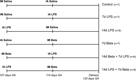
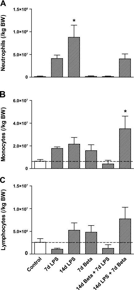
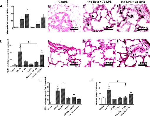
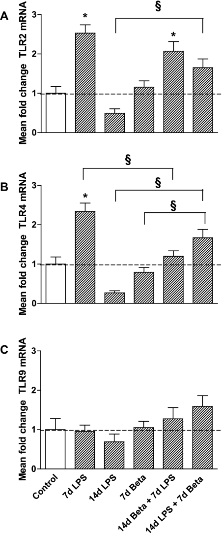
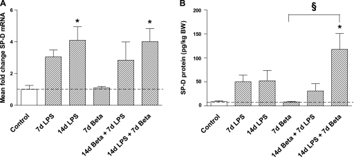
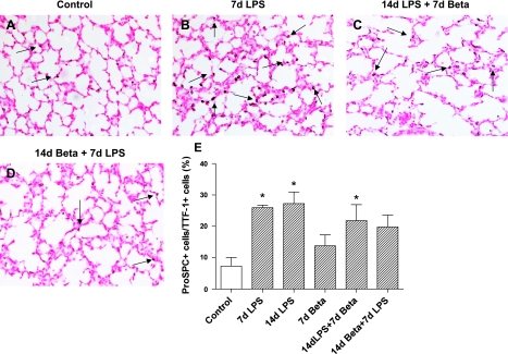
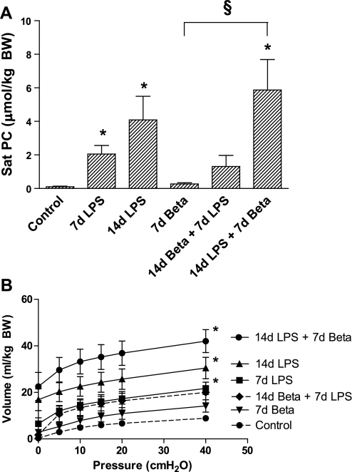
The proinflammatory stimulus of chorioamnionitis is commonly associated with preterm delivery. Women at risk of preterm delivery receive antenatal glucocorticoids to functionally mature the fetal lung. However, the effects of the combined exposures of chorioamnionitis and antenatal glucocorticoids on the fetus are poorly understood. Time-mated ewes with singleton fetuses received an intra-amniotic injection of lipopolysaccharide (LPS) either preceding or following maternal intramuscular betamethasone 7 or 14 days before delivery, and the fetuses were delivered at 120 days gestational age (GA) (term = 150 days GA). Gestation matched controls received intra-amniotic and maternal intramuscular saline. Compared with saline controls, intra-amniotic LPS increased inflammatory cells in the bronchoalveolar lavage and myeloperoxidase, Toll-like receptor 2 and 4 mRNA, PU.1, CD3, and Foxp3-positive cells in the fetal lung. LPS-induced lung maturation measured as increased airway surfactant and improved lung gas volumes. Intra-amniotic LPS-induced inflammation persisted until 14 days after exposure. Betamethasone treatment alone induced modest lung maturation but, when administered before intra-amniotic LPS, suppressed lung inflammation. Interestingly, betamethasone treatment after LPS did not counteract inflammation but enhanced lung maturation. We conclude that the order of exposures of intra-amniotic LPS or maternal betamethasone had large effects on fetal lung inflammation and maturation.


Figures

Fig. 1.
Fig. 1.
Study design. Pregnant ewes received an intra-amniotic (IA) injection of lipopolysaccharide (LPS) and/or an intramuscular (IM) injection of betamethasone (Beta) and/or an equivalent injection of saline for control animals at 107 days and/or 114 days gestation (GA). Lambs were delivered preterm by cesarean section at 120 days GA (term = 150 days GA). 7d, 7-day exposure; 14d, 14-day exposure.
Fig. 2.
Fig. 2.
Differential cell count of the bronchoalveolar lavage. A: neutrophil levels increased 14 days after the exposure to LPS. B: combined exposure to LPS for 14 days and betamethasone for 7 days increased monocytes in the bronchoalveolar lavage. C: lymphocyte count did not differ in any of the treatment groups compared with controls. *P < 0.05 vs. controls by 1-way ANOVA with Tukey's post hoc test. BW, body weight.
Fig. 3.
Fig. 3.
Characterization of inflammatory cells in the fetal lung tissue. A: quantitation of myeloperoxidase (MPO)-expressing cells in lung sections per microscopic field corrected for the tissue fraction (see methods for details). Representative photomicrographs of immunostaining against myeloperoxidase using lung sections from controls (B), 14d betamethasone + 7d LPS (C), and 14d LPS + 7d betamethasone (D). MPO-positive cells increased in the lung tissue 14 days after LPS exposure compared with controls. E: quantitation of PU.1 expressing cells in lung sections per microscopic field. Representative photomicrographs of immunostaining against PU.1 using lung sections from controls (F), 14d betamethasone + 7d LPS (G), and 14d LPS + 7d betamethasone (H). LPS exposure for 7 days and combined exposure to LPS for 14 days and betamethasone for 7 days increased the number of PU.1-positive cells in the fetal lung. Betamethasone treatment 7 days before the LPS exposure prevented this increase. I: number of CD3-positive cells increased significantly 7 and 14 days after LPS exposure compared with controls. J: Foxp3 expression increased 7 days after LPS exposure compared with controls. Betamethasone treatment before the LPS exposure prevented this increase. *P < 0.05 vs. controls; §P < 0.05 between experimental groups by 1-way ANOVA with Tukey's post hoc test.
Fig. 4.
Fig. 4.
Expression of Toll-like receptors (TLR) 2, 4, and 9. TLR2 (A) and TLR4 (B) mRNA expression were upregulated 7 days after LPS exposure but returned to baseline 14 days after exposure to LPS. Betamethasone treatment before LPS exposure prevented increased TLR4 expression, but not TLR2 expression. TLR2 and TLR4 mRNA levels increased in the lungs of lambs that received betamethasone after LPS exposure, although these levels were not higher than levels measured in controls. C: TLR9 was not differently expressed in experimental groups compared with controls. *P < 0.05 vs. controls; §P < 0.05 between experimental groups by 1-way ANOVA with Tukey's post hoc test.
Fig. 5.
Fig. 5.
Pulmonary surfactant protein D (SP-D) mRNA and protein expression. A: SP-D mRNA levels increased 14 days after the exposure to LPS irrespectively of the betamethasone posttreatment. B: only combined exposure to LPS for 14 days and betamethasone for 7 days increased SP-D protein expression in the bronchoalveolar lavage fluid. Exposure to LPS for 14 days did not increase SP-D protein expression. *P < 0.05 vs. controls; §P < 0.05 between experimental groups by 1-way ANOVA with Tukey's post hoc test.
Fig. 6.
Fig. 6.
Pro-pulmonary surfactant protein C (pro-SP-C) immunostaining in the lung. The number of pro-SP-C-positive cells was expressed as a percentage of TTF-1-positive cells (data in Table 4). Representative photomicrographs of immunostaining using lung sections from controls (A), 7d LPS (B), 14d LPS + 7d betamethasone (C), and 14d betamethasone + 7d LPS (D). E: quantitation of pro-SP-C-expressing cells in lung sections per microscopic field via a ×20 objective. LPS exposure increased pro-SP-C expression in the alveolar type II cells in the lung. (*P < 0.05 vs. controls, scale bar is 50 μm).
Fig. 7.
Fig. 7.
Saturated phosphatidylcholine (Sat PC) and pressure-volume curves. A: Sat PC levels in the bronchoalveolar lavage fluid were increased 7 and 14 days after exposure to intra-amniotic LPS. Betamethasone posttreatment 7 days after the exposure to LPS increased Sat PC levels further. B: pressure-volume curve of animals after 14 days of LPS exposure was significantly higher compared with controls irrespectively of treatment with betamethasone. *P < 0.05 vs. controls; §P < 0.05 between experimental groups by 1-way ANOVA with Tukey's post hoc test.

Similar articles

Cited by

References

    1. Andrews WW, Goldenberg RL, Faye-Petersen O, Cliver S, Goepfert AR, Hauth JC. The Alabama Preterm Birth study: polymorphonuclear and mononuclear cell placental infiltrations, other markers of inflammation, and outcomes in 23- to 32-week preterm newborn infants. Am J Obstet Gynecol 195: 803–808, 2006 - PubMed
    1. Bachurski CJ, Ross GF, Ikegami M, Kramer BW, Jobe AH. Intra-amniotic endotoxin increases pulmonary surfactant proteins and induces SP-B processing in fetal sheep. Am J Physiol Lung Cell Mol Physiol 280: L279–L285, 2001 - PubMed
    1. Ballard PL, Ertsey R, Gonzales LW, Gonzales J. Transcriptional regulation of human pulmonary surfactant proteins SP-B and SP-C by glucocorticoids. Am J Respir Cell Mol Biol 14: 599–607, 1996 - PubMed
    1. Ballard PL, Granberg P, Ballard RA. Glucocorticoid levels in maternal and cord serum after prenatal betamethasone therapy to prevent respiratory distress syndrome. J Clin Invest 56: 1548–1554, 1975 - PMC - PubMed
    1. Banks BA, Cnaan A, Morgan MA, Parer JT, Merrill JD, Ballard PL, Ballard RA. Multiple courses of antenatal corticosteroids and outcome of premature neonates. North American Thyrotropin-Releasing Hormone Study Group. Am J Obstet Gynecol 181: 709–717, 1999 - PubMed

Publication types

MeSH terms