. 2012 Feb;23(3):464-79.
doi: 10.1091/mbc.E11-07-0590. Epub 2011 Dec 7.

Promiscuous targeting of polytopic membrane proteins to SecYEG or YidC by the Escherichia coli signal recognition particle

Affiliations

Promiscuous targeting of polytopic membrane proteins to SecYEG or YidC by the Escherichia coli signal recognition particle

Thomas Welte et al. Mol Biol Cell. 2012 Feb.

Abstract

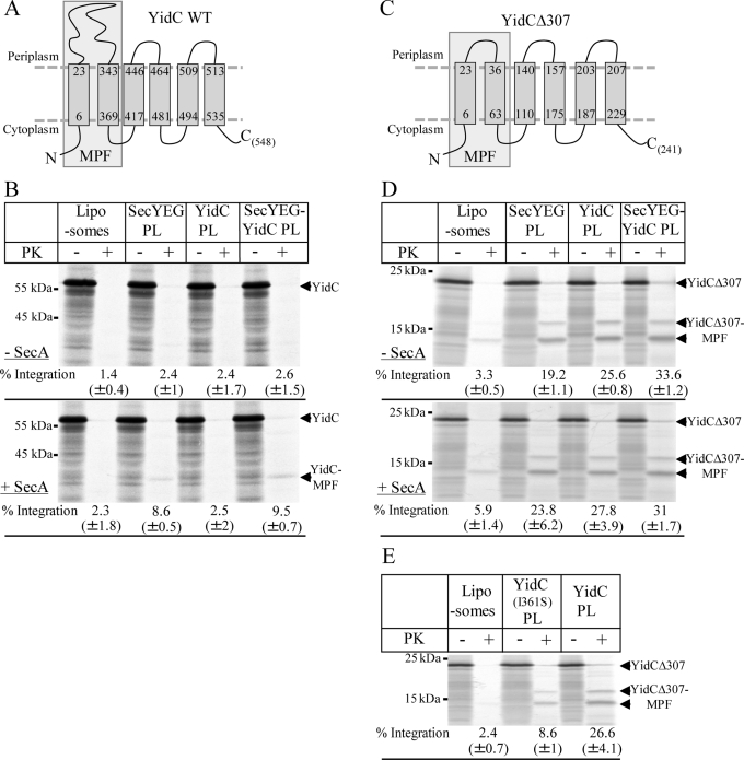
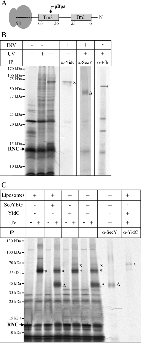
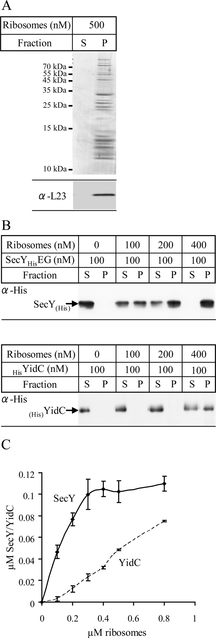
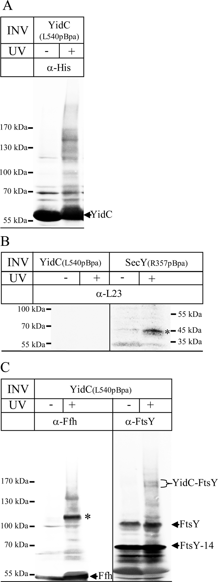
Protein insertion into the bacterial inner membrane is facilitated by SecYEG or YidC. Although SecYEG most likely constitutes the major integration site, small membrane proteins have been shown to integrate via YidC. We show that YidC can also integrate multispanning membrane proteins such as mannitol permease or TatC, which had been considered to be exclusively integrated by SecYEG. Only SecA-dependent multispanning membrane proteins strictly require SecYEG for integration, which suggests that SecA can only interact with the SecYEG translocon, but not with the YidC insertase. Targeting of multispanning membrane proteins to YidC is mediated by signal recognition particle (SRP), and we show by site-directed cross-linking that the C-terminus of YidC is in contact with SRP, the SRP receptor, and ribosomal proteins. These findings indicate that SRP recognizes membrane proteins independent of the downstream integration site and that many membrane proteins can probably use either SecYEG or YidC for integration. Because protein synthesis is much slower than protein transport, the use of YidC as an additional integration site for multispanning membrane proteins may prevent a situation in which the majority of SecYEG complexes are occupied by translating ribosomes during cotranslational insertion, impeding the translocation of secretory proteins.


Figures

FIGURE 1:
FIGURE 1:
Reconstitution of SecYEG and YidC into proteoliposomes. (A) Proteoliposomes were prepared using E. coli phospholipids supplemented with 5% DAG. Purified SecYEG or YidC were reconstituted into liposomes and pelleted by centrifugation. The pellet was resuspended and adjusted to a final concentration of 0.4 mg/ml SecY, 0.4 mg/ml YidC, or 0.4 mg/ml SecY + 0.4 mg/ml YidC. One aliquot (4 μl) was loaded onto a 15% SDS gel and Coomassie stained. Pure liposomes served as control. (B) The proteoliposomes (2 μl) shown in A were separated on SDS–PAGE and blotted onto a nitrocellulose membrane. The membrane was subsequently cut into two pieces, and the upper part was decorated with α-YidC antibodies and the lower part with α-SecY antibodies. (C) One aliquot of the purified E. coli in vitro TT system used for in vitro protein synthesis was probed with the indicated antibodies after Western blotting. Purified FtsY (0.7 μg), Ffh (0.2 μg), SecA (1.5 μg), SecY (0.8 μg), and YidC (0.8 μg) served as controls.
FIGURE 2:
FIGURE 2:
Integration of in vitro–synthesized TatC into inner membrane vesicles. (A) Predicted topology of TatC according to Behrendt et al. (2004); a His10 tag was fused to its N-terminus. The boxed portion of TatC most likely corresponds to the 19-kDa, membrane-protected fragment of TatC (TatC-MPF). (B) 35S-Labeled His10-TatC was in vitro synthesized in the absence (–INV) or presence of wild-type E. coli inner membrane vesicles (WT INV; 2 mg/ml). After synthesis, one-fourth of the reaction was precipitated with TCA, and the remainder was first treated with 0.5 mg/ml PK for 30 min at 25°C and then TCA precipitated. Full-size TatC (TatC) and the TatC-MPF are indicated. Note that wild-type E. coli INV contains sufficient amounts of SRP and FtsY (Koch et al., 1999). The percentages of PK protection was calculated using ImageQuant (GE Healthcare) by quantifying the ratio of radioactivity present in the PK-treated sample and the directly TCA-precipitated sample and are the mean values of at least three independent experiments. Note that the calculation is corrected for the loss of methionine/cysteine residues and based on the assumption that TatC-MPF corresponds to the first four TMs of TatC. (C) TatC was in vitro synthesized as in B but extracted with alkaline Na2CO3 (pH 11.3, 0.2 M final concentration). After ultracentrifugation, pellet (P) and supernatant (S) were separated by SDS–PAGE. For quantification, the amounts of radioactive material in both fractions were set as 100%, and the distribution between both fractions was calculated. The values provided are the mean values of at least three independent experiments, and the SD is indicated.
FIGURE 3:
FIGURE 3:
TatC is targeted by SRP to either SecYEG or YidC. (A) 35S-Labeled His10-TatC was in vitro synthesized as described in Figure 1. When indicated, SRP (Ffh [150 nM] + 4.5S RNA [15 μg/ml]) and FtsY (750 nM) were present during synthesis. Synthesis was performed in the presence of 0.4 mg/ml of the liposomes or proteoliposomes (PL) shown in Figure 1. After synthesis, the samples were PK digested. (B) Carbonate resistance of TatC synthesized in the presence of liposomes or proteoliposomes. The mean values of three independent experiments are shown. (C) 35S-Labeled His10-TatC and 35S-labeled TatC were synthesized in the presence of SRP, FtsY, and proteoliposomes reconstituted with either wild-type YidC or the YidC(I361S) mutant. (D) Coomassie blue staining and immunodetection of proteoliposomes containing either wild-type YidC or the YidC(I361S) mutant. Four μl of each proteoliposome preparation were loaded for Coomassie staining and 2 μl for immunodetection.
FIGURE 4:
FIGURE 4:
YidC is sufficient for MtlA integration. (A) Topology of MtlA according to Sugiyama et al. (1991); the cleavage site of PK is indicated. The MPF corresponds to the first six TMs. (B) 35S-Labeled MtlA was synthesized in the presence (WT INV) or absence (–INV) of INV. After synthesis, one-half of the reaction was precipitated with TCA, and the other half of the reaction was first treated with 0.5 mg/ml PK for 30 min at 25°C and then TCA precipitated. Full-length MtlA and the membrane-protected fragment (MtlA-MPF) corresponding to the hydrophobic core of MtlA are indicated. The band labeled with an asterisk probably corresponds to a product of premature termination of protein synthesis. (C) MtlA was synthesized as described in B and under the same conditions as described in the legend to Figure 3A. (D) MtlA was synthesized as described in Figure 3C.
FIGURE 5:
FIGURE 5:
Membrane protein integration via YidC is restricted to membrane proteins with small periplasmic loops. (A) Predicted topology of YidC. The cleavage site for PK is indicated. The YidC-MPF corresponds to the first two TMs and the connecting periplasmic loop (Koch et al., 2002). (B) YidC was synthesized in presence of SRP, FtsY, and liposomes/proteoliposomes as indicated. Protease protection was analyzed in the absence or presence of 600 nM SecA. Full-length YidC and YidC-MPFs are indicated. (C) Predicted topology of YidCΔ307 (Deitermann et al., 2005). (D) Protease protection of YidCΔ307. The lower, 13-kDa MPF was used for calculating integration rates. (E) Protease protection of YidCΔ307 in the presence of proteoliposomes containing wild-type YidC or the YidC(I361S) mutant.
FIGURE 6:
FIGURE 6:
Molecular contacts of YidCΔ307 RNCs in INV and proteoliposomes. (A) Cartoon showing the 98–amino acid long YidCΔ307-RNCs used in this study. YidCΔ307-98 RNCs comprise the first 98 amino acids of YidCΔ307 (Figure 5C) and contains pBpa at position 46 in TM 2. (B) YidCΔ307-98 RNCs were synthesized in the presence of INVs. Cross-linking products were identified by immunoprecipitations after separation on 15% SDS–PAGE. (C) YidCΔ307-RNCs were synthesized in the presence of SRP, purified via a sucrose cushion, and incubated with FtsY and liposomes/proteoliposomes as indicated. Samples were loaded on a 7–18% SDS gel. Cross-links to Ffh (asterisk), YidC (cross), and SecY (triangle) are indicated.
FIGURE 7:
FIGURE 7:
YidC binds to E. coli ribosomes. (A) Purified and salt-washed 70S ribosomes (500 nM) were incubated in binding buffer (pH 7.6). After 30 min of incubation on ice, ribosomes were centrifuged through a sucrose cushion. Pellet (P) and supernatant fractions (S) were separated on SDS gels and analyzed by Coomassie staining (top) and immunodetection using antibodies against L23 (bottom). (B) Increasing concentrations of salt-washed ribosomes were incubated with either 100 nM detergent-purified SecYEG (top) or detergent-purified YidC (bottom) in binding buffer (pH 7.6) and subjected to the same conditions as described in A. Pellet and supernatant fractions were immunodetected using antibodies against the N-terminal His tags of SecY or YidC. The percentage of binding was calculated using ImageJ software. (C) Quantification of at least three independent ribosome-binding experiments, using varying concentrations of SecYEG or YidC, respectively. The amount of ribosome-bound SecY/YidC was plotted against the total ribosome concentration.
FIGURE 8:
FIGURE 8:
In vitro cross-links using INVs of YidC pBpa mutants show contacts to SRP and FtsY. (A) One mg of INVs from E. coli cells overexpressing YidC(L540pBpa) was resuspended in 250 μl of INV buffer and cross-linked by UV irradiation. The samples were separated on a 5–15% SDS gel, and immunodetection was performed with α-His antibodies. (B) Cross-linking with INVs purified from E. coli cells expressing YidC(L540pBpa) or the pBpa-containing SecY derivative SecY(R357pBpa), which contains pBpa within the fifth cytosolic loop of SecY. After separation on SDS–PAGE, cross-links to L23 (asterisk) were identified by immunodetection. (C) The material shown in A was also probed with α-Ffh and α-FtsY antibodies. The YidC-Ffh cross-link at 110 kDa is indicated (asterisk), as well as the Ffh that was present in the INVs. The YidC-FtsY cross-link appears in two distinct bands at ∼160 kDa, which probably reflect the two FtsY isoforms present in E. coli INV. FtsY corresponds to full-size FtsY, whereas FtsY-14 corresponds to an N-terminally–truncated FtsY derivative.

Similar articles

Cited by

References

    1. Ahrem B, Hoffschulte HK, Muller M. In vitro membrane assembly of a polytopic, transmembrane protein results in an enzymatically active conformation. J Cell Biol. 1989;108:1637–1646. - PMC - PubMed
    1. Baars L, Wagner S, Wickstrom D, Klepsch M, Ytterberg AJ, van Wijk KJ, de Gier JW. Effects of SecE depletion on the inner and outer membrane proteomes of Escherichia coli. J Bacteriol. 2008;190:3505–3525. - PMC - PubMed
    1. Beck K, Eisner G, Trescher D, Dalbey RE, Brunner J, Muller M. YidC, an assembly site for polytopic Escherichia coli membrane proteins located in immediate proximity to the SecYE translocon and lipids. EMBO Rep. 2001;2:709–714. - PMC - PubMed
    1. Beck K, Wu LF, Brunner J, Muller M. Discrimination between SRP- and SecA/SecB-dependent substrates involves selective recognition of nascent chains by SRP and trigger factor. EMBO J. 2000;19:134–143. - PMC - PubMed
    1. Beha D, Deitermann S, Muller M, Koch HG. Export of beta-lactamase is independent of the signal recognition particle. J Biol Chem. 2003;278:22161–22167. - PubMed

Publication types

MeSH terms

Substances

LinkOut - more resources