. 2011 Dec 27;108(52):21046-51.
doi: 10.1073/pnas.1112715108. Epub 2011 Dec 12.

Structural interconversions modulate activity of Escherichia coli ribonucleotide reductase

Affiliations

Structural interconversions modulate activity of Escherichia coli ribonucleotide reductase

Nozomi Ando et al. Proc Natl Acad Sci U S A. .

Abstract

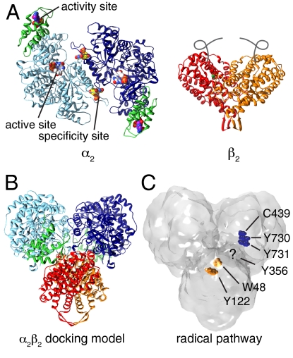
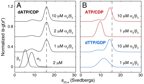
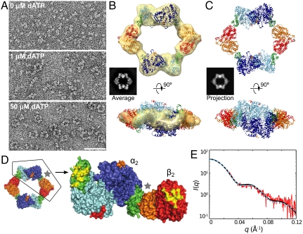
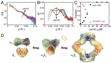
Essential for DNA biosynthesis and repair, ribonucleotide reductases (RNRs) convert ribonucleotides to deoxyribonucleotides via radical-based chemistry. Although long known that allosteric regulation of RNR activity is vital for cell health, the molecular basis of this regulation has been enigmatic, largely due to a lack of structural information about how the catalytic subunit (α(2)) and the radical-generation subunit (β(2)) interact. Here we present the first structure of a complex between α(2) and β(2) subunits for the prototypic RNR from Escherichia coli. Using four techniques (small-angle X-ray scattering, X-ray crystallography, electron microscopy, and analytical ultracentrifugation), we describe an unprecedented α(4)β(4) ring-like structure in the presence of the negative activity effector dATP and provide structural support for an active α(2)β(2) configuration. We demonstrate that, under physiological conditions, E. coli RNR exists as a mixture of transient α(2)β(2) and α(4)β(4) species whose distributions are modulated by allosteric effectors. We further show that this interconversion between α(2)β(2) and α(4)β(4) entails dramatic subunit rearrangements, providing a stunning molecular explanation for the allosteric regulation of RNR activity in E. coli.


Conflict of interest statement

The authors declare no conflict of interest.

Figures

Fig. 1.
Fig. 1.
Previously determined structures and proposed models for E. coli class Ia RNR. (A) Homodimeric α2 with nucleotides bound (spheres) and the cone domains (residues 1–100) colored in green. Homodimeric β2 with diiron centers (green) and disordered C termini (gray lines). Protein Data Bank ID codes 4R1R, 3R1R, 1RIB (5, 15, 28). (B) Proposed α2β2 model in which the subunits are docked along their symmetry axis (15). (C) Docking model rendered as a surface. Radical pathway involving Y122 → W48 → Y356 in β2 (Y122 and W48 shown as orange spheres) and residues Y731 → Y730 → C439 in α2 (dark-blue spheres). Y356 in β2 lies in the disordered C termini (15).
Fig. 2.
Fig. 2.
Sedimentation coefficient distributions of E. coli class Ia RNR: dATP and high protein concentrations shift the equilibrium toward a large complex. Physiologically relevant effector and substrate concentrations were chosen to saturate their respective sites, based on previously reported nucleotide-binding affinities (4, 24, 25, 34, 51). (A) In the presence of 175 μM dATP and 1 mM CDP, the individual subunits (each at 2 μM) sediment at 5.2 and 8.4 S. When mixed, a single slowly dissociating 15.6 S complex is observed with a molecular mass of 533 kDa. (B) In the presence of 3 mM ATP and 1 mM CDP (red curves) or 0.1 mM dTTP and 1 mM GDP (blue curves), broad, protein concentration-dependent peaks are observed, indicative of multiple species in rapid exchange. Raising the protein concentration leads to a peak shift toward 15.6 S (position indicated by dashed line). Curves are offset by a constant value for clarity.
Fig. 3.
Fig. 3.
Structure of the dATP α4β4 complex by EM and X-ray crystallography. (A) EM images of RNR in the presence of 1 mM CDP and increasing dATP concentrations show the formation of α4β4 rings. (B) A class average with 1039 particles (Average) is representative of the ring structures observed at 50 μM dATP and is composed of alternating α2 and β2 subunits as indicated by its close resemblance to the 2D projection of the α4β4 crystal structure (Projection in C). These insets are 314-Å wide. Crystal structures of individual α2 and β2 subunits (5, 39), colored as in Fig. 1, fit to a 3D EM map of the α4β4 ring structure. (C) Crystal structure of dATP-bound RNR at 5.65-Å resolution with the asymmetric unit containing an α4β4 ring that agrees well with the EM model (5–9 Å Cα rmsd). (D) Surface rendering of the crystal structure with half of the ring removed, revealing the areas on α and β that are buried (yellow) upon formation of the α4β4 ring. (E) Experimental solution scattering (red) of 2 μM RNR in the presence of saturating dATP/CDP superimposed with the theoretical scattering curves calculated from the EM model (black solid) and crystal structure (cyan dashed).
Fig. 4.
Fig. 4.
SAXS investigation of the transition between lower and higher order structures of E. coli class Ia RNR. (A) Scattering curves measured as 0–175 μM dATP was titrated into a 6 μM solution of α2 and β2 in the presence of 1 mM CDP (red to violet) display isointensity points, suggesting a two-state transition. (B) Kratky representations of the scattering curves (Iq2 vs. q) at 0 μM (red), 12 μM (green), and 40 μM (blue) dATP show a transition from a compact globular state, as indicated by the monomodal peak, to a large nonglobular state, indicated by a bimodal curve (36). (C) Fitting linear combinations of the α2β2 docking model (15) and the α4β4 ring to the titration data provided relative fractions of the two states. (D) Ab initio SAXS reconstructions of free subunits aligned with deposited crystal structures (5, 39), a compact α2β2 state aligned with the proposed docking model (15), and the α4β4 ring aligned with the crystal structure (SI Appendix, Tables S2 and S3, and Fig. S10). The small additional density observed in the molecular envelope of α2β2 can be explained by the presence of approximately 3% α4β4 (Fig. 4C).

References

    1. Licht S, Gerfen GJ, Stubbe J. Thiyl radicals in ribonucleotide reductases. Science. 1996;271:477–481. - PubMed
    1. Jordan A, Reichard P. Ribonucleotide reductases. Annu Rev Biochem. 1998;67:71–98. - PubMed
    1. Nordlund P, Reichard P. Ribonucleotide reductases. Annu Rev Biochem. 2006;75:681–706. - PubMed
    1. Brown NC, Reichard P. Role of effector binding in allosteric control of ribonucleoside diphosphate reductase. J Mol Biol. 1969;46:39–55. - PubMed
    1. Eriksson M, et al. Binding of allosteric effectors to ribonucleotide reductase protein R1: Reduction of active-site cysteines promotes substrate binding. Structure. 1997;5:1077–1092. - PubMed

Publication types

Associated data

LinkOut - more resources