. 2011 Dec 27;108(52):21093-8.
doi: 10.1073/pnas.1112061109. Epub 2011 Dec 12.

Neuronal differentiation by TAp73 is mediated by microRNA-34a regulation of synaptic protein targets

Affiliations

Neuronal differentiation by TAp73 is mediated by microRNA-34a regulation of synaptic protein targets

Massimiliano Agostini et al. Proc Natl Acad Sci U S A. .

Abstract

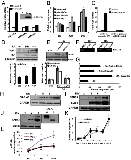
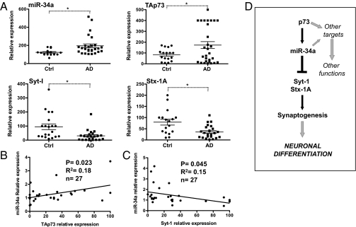
The p53-family member TAp73 is a transcription factor that plays a key role in many biological processes. Here, we show that p73 drives the expression of microRNA (miR)-34a, but not miR-34b and -c, by acting on specific binding sites on the miR-34a promoter. Expression of miR-34a is modulated in parallel with that of TAp73 during in vitro differentiation of neuroblastoma cells and cortical neurons. Retinoid-driven neuroblastoma differentiation is inhibited by knockdown of either p73 or miR-34a. Transcript expression of miR-34a is significantly reduced in vivo both in the cortex and hippocampus of p73(-/-) mice; miR-34a and TAp73 expression also increase during postnatal development of the brain and cerebellum when synaptogenesis occurs. Accordingly, overexpression or silencing of miR-34a inversely modulates expression of synaptic targets, including synaptotagmin-1 and syntaxin-1A. Notably, the axis TAp73/miR-34a/synaptotagmin-1 is conserved in brains from Alzheimer's patients. These data reinforce a role for TAp73 in neuronal development.


Conflict of interest statement

The authors declare no conflict of interest.

Figures

Fig. 1.
Fig. 1.
TAp73 drives the expression of miR-34a and in vitro terminal differentiation of cortical neurons. (A) SAOS-2-TAp73β inducible cell lines were treated with Doxycyclin for 24 h to overexpress the human TAp73β protein (Inset, Western blot). Endogenous levels of miRs-34a, -34b, and -34c were assessed by real-time PCR. (B) SAOS-2-TAp73β inducible cell lines were treated with Doxycyclin for the indicate times and endogenous levels of pri-miR-34a, pre-miR-34a, and miR-34 were evaluated by real-time PCR. *P < 0.05. (C) The miR-34a promoter region between −1,472 and +551 bp, which contains a p53 consensus site was analyzed for TAp73β responsiveness. Insertion of this miR-34a promoter region in a luciferase reporter gene leads to increased luciferase activity in the presence of TAp73β in SAOS-2 cells. (D) TAp73 regulates miR-34 in RA-induced terminal differentiation of a neuroblastoma cell line. SHSY-5Y cells were treated with 10 μM RA and expression of TAp73, p21, and β-tubulin was assessed by Western blot. Expression of miR-34a was evaluated by real-time PCR (Lower, histogram). (E) p73siRNA prevents the up-regulation of precursor-miR-34a and miR-34a after RA treatment. *P < 0.05. (F) ChIP assay on SHSY-5Y cells untreated (ctrl) or treated with RA. Protein-chromatin complexes were immunoprecipitated with anti-TAp73 or IgG as control antibody. RT-PCR was performed with primers designed against the hsa-miR-34a promoter region containing the predicted and validated p53 binding site. (G) Neurite outgrowth in SHSY-5Y untransfected (ctrl), or transfected with scrambled, siRNAp73, or anti-miR-34a and then treated with RA for 48 h. *P < 0.05. (H and I) Western blot analysis of three different neuronal markers and GAPDH in cultured cortical neurons at DIV2, -5, -6, and -7. Syn II (Synapsin II). (J and K) Cortical neurons were collected at the indicated time points and expression of miR-34a and TAp73 was evaluated by Western blot and real-time PCR respectively. Data represent mean ± SD of three different experiments. *P = 0.016. (L) Relative expression of miR-34a compared between WT, TAp73−/−, and p73−/− mice during terminal neuronal differentiation. Data are normalized to the housekeeping gene Sno202 relative to DIV2. Data represent mean ± SD of three different experiments. *P = 0.045.
Fig. 2.
Fig. 2.
Expression of miR-34a and TAp73 in vivo. (A) Expression of miR-34a is reduced in p73−/− mice. Cortex and hippocampus were isolated from p73+/+, p73+/−, and p73−/− mice (n = 5–9; age, P2), and levels of miR-34a were evaluated by real-time PCR. **P < 0.01. (B) Different regions of the cortex and hippocampus were isolated by laser-capture microdissection from p73+/+ and p73−/− mice and levels of miR-34a were evaluated by real-time PCR. Data represent mean ± SD of three different experiments. *P < 0.05. (C) The cerebellum was isolated from WT mice (n = 4 per each point) and expression of miR-34a and TAp73 was evaluated during cerebellar development by real-time PCR at birth (P0) and subsequent postnatal days. (D) Relative expression of miR-34a in the cerebellum from p73+/+ and p73−/− mice during development. Data are normalized to the housekeeping gene Sno202 relative to P1. Data represent mean ± SD of three different experiments. *P = 0.045.
Fig. 3.
Fig. 3.
miR-34a modulates the expression of synaptic markers. (A) WT cortical neurons (DIV3) were transfected with pre-miR-34a or a scrambled control (Scramble) and after 24 h, mRNA levels of several neuronal markers were determined by real-time PCR. Stx (syntaxin), Syn (synapsin), Syt (synaptotagmin). Data represent mean ± SD of three different experiments. *P = 0.01. (B) Insertion of Syt-I 3′ UTR or Stx1A 3′ UTR (WT or deleted) upstream of a luciferase reporter gene shows diminished luciferase activity in the presence precursor miR-34a. The assay was performed in HEK 293E cells. Data represent mean ± SD of three different experiments. *P = 0.03. (C) Cortical neurons were transfected with precursor miR-34a (Pre-34a) or scrambled control (Scramble) for 72 h and the cell lysates were analyzed for Syt-I and Stx-1A expression. (D) WT cortical neurons (DIV3) were transfected with anti–miR-34a or scrambled control (Scramble) and, after 24 h, mRNA levels of the indicated synaptic proteins were determined by real-time PCR. Data represent mean SD of three different experiments. *P = 0.03. (E) Western blot analysis of proteins from cortical neurons transfected at DIV2 with anti–miR-34a or scrambled control. Actin or GAPDH were used as loading controls. Numbers below the Western blot indicate the relative levels of synaptic protein normalized to actin or GAPDH related to the scrambled control. (F) The indicated cell layers of the cerebellum were isolated by laser capture microdissection (Fig. S7) and levels of miR-34a and Syt-I were evaluated by real-time PCR. Data are normalized to the housekeeping gene Sno202 or GAPDH relative to granular cell layer expression. Data represent mean ± SD of three different experiments.
Fig. 4.
Fig. 4.
Expression of miR-34a, TAp73, and synaptic proteins in AD brain. (A) Total RNA was extracted from postmortem control and AD hippocampus. Expression of the indicated genes was evaluated by real-time PCR. Data represent mean ± SEM. *P < 0.05. (B) Pearson's test on individual AD samples show significant positive correlation between TAp73 and miR-34a expression. (C) A significant negative correlation between miR-34a and Syt-1. (D) Schematic model for the role of TAp73/miR-34a axis in developmental neuronal differentiation.

References

    1. Müller M, et al. TAp73/Delta Np73 influences apoptotic response, chemosensitivity and prognosis in hepatocellular carcinoma. Cell Death Differ. 2005;12:1564–1577. - PubMed
    1. Wang J, et al. TAp73 is a downstream target of p53 in controlling the cellular defense against stress. J Biol Chem. 2007;282:29152–29162. - PubMed
    1. Grob T-J, et al. Human delta Np73 regulates a dominant negative feedback loop for TAp73 and p53. Cell Death Differ. 2001;8:1213–1223. - PubMed
    1. Yang A, et al. p73-deficient mice have neurological, pheromonal and inflammatory defects but lack spontaneous tumours. Nature. 2000;404:99–103. - PubMed
    1. De Laurenzi V, et al. Induction of neuronal differentiation by p73 in a neuroblastoma cell line. J Biol Chem. 2000;275:15226–15231. - PubMed

Publication types

MeSH terms