. 2011 Dec 27;108(52):21099-104.
doi: 10.1073/pnas.1112063108. Epub 2011 Dec 12.

microRNA-34a regulates neurite outgrowth, spinal morphology, and function

Affiliations

microRNA-34a regulates neurite outgrowth, spinal morphology, and function

Massimiliano Agostini et al. Proc Natl Acad Sci U S A. .

Abstract

The p53 family member TAp73 is a transcription factor that plays a key role in many biological processes, including neuronal development. In particular, we have shown that p73 drives the expression of miR-34a, but not miR-34b and c, in mouse cortical neurons. miR-34a in turn modulates the expression of synaptic targets including synaptotagmin-1 and syntaxin-1A. Here we show that this axis is retained in mouse ES cells committed to differentiate toward a neurological phenotype. Moreover, overexpression of miR-34a alters hippocampal spinal morphology, and results in electrophysiological changes consistent with a reduction in spinal function. Therefore, the TAp73/miR-34a axis has functional relevance in primary neurons. These data reinforce a role for miR-34a in neuronal development.


Conflict of interest statement

The authors declare no conflict of interest.

Figures
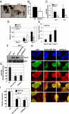
Fig. 1.
Fig. 1.
The TAp73/miR-34a pathway participates in neuronal commitment of murine ES cells. (A) Hippocampal morphology is altered in the DG of p73−/− mice. Golgi staining of DG from p73+/+ and p73−/− mice (age P18). Brain coronal sections were treated as described in Methods. A representative photomicrograph is shown. (B) Proliferating cells are reduced in the SGZ of miR-34a−/− mice. Graph shows the mean ± SD of Ki-67–positive cells. (C) miR-34a expression is reduced in the hippocampus of p73−/− mice between E17 and postnatal day 3. Hippocampus was isolated from p73+/+ and p73−/− mice (n = 3), and levels of miR-34a were evaluated by real-time PCR (*P < 0.05). (D and E) Murine ES cells were cultivated on fixed feeder NIH 3T3 cells in the absence of serum, as described in Methods. Differentiated cells were collected at the indicated times and neural differentiation was evaluated. Real-time PCR analysis demonstrated that ES cells rapidly and efficiently differentiated into neural cells, as indicated by the induction of neural markers, such as neurofilament (NeuF) and βIII-tubulin (βIII-Tub). At the indicated times, RNA extractions were prepared for real-time PCR analysis of βIII-tubulin, neurofilament, TAp73, and miR-34a. Neural differentiation was evident at the RNA level by elevation of the putative neural markers βIII-tubulin and neurofilament. Expression of p73 and miR34a was also enhanced in parallel during differentiation. (H) Immunofluorescent staining was performed at day 7 of differentiation, showing typical neurite formation in control cells (Ctrl) that were also immunoreactive with Pax6 and βIII-tubulin antibodies. Merge of the same fields is shown, and higher magnification of the merged image is also shown (Insets). The same analysis was performed in cells transfected with anti–miR-34a or sip73 (F and G), and, in both cases, shows a reduction in Pax6 and βIII-tubulin immunoreactivity, together with reductions in neurite outgrowth. In the same experiment, changes in p73, neurofilament, and βIII-tubulin (D) and miR-34a (E) expression were examined at days 4 and 7. Change in expression of TAp73 and miR-34a at days 0 and 7 in control cells (Ctrl) and cells transfected with anti–miR-34a (D) or siRNAp73 (E) are shown along with corresponding changes in expression of βIII-tubulin and neurofilament at day 7 (I).
Fig. 2.
Fig. 2.
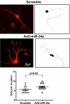
miR-34a negatively affects dendritic outgrowth of cortical neurons. DIV 2 WT cortical neurons were transfected with GFP plus scramble or GFP plus anti–miR-34a (100 nM). After 72 h, neurons were fixed and mounted for confocal microscopy. A representative image is shown (Left). Right: Tracing of projections of the dendritic tree of one representative neuron. Quantification of branch number after inhibition of miR-34a was performed as described in Methods. In each experiment, nine to 15 cells were analyzed. Data represent mean ± SD of three different experiments (*P = 0.02, two-tailed Student t test).
Fig. 3.
Fig. 3.
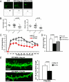
miR-34a negatively affects dendritic outgrowth of cortical neurons. (A) DIV 2 WT cortical neurons were transfected with GFP, GFP plus scrambled control (neg ctrl), or GFP plus pre-34a (30 nM). After 72 h, neurons were fixed and mounted for confocal microscopy. Representative images are shown (Upper). Lower: Tracings of projections of the dendritic tree of one representative neuron. (B) Quantification of total length and branch number after ectopic expression of miR-34a was performed as described in Methods. (C) Dendritic complexity of cortical neurons transfected as in B was evaluated by using Sholl analysis. In each experiment, nine to 15 cells were analyzed. Data represent mean ± SD of three different experiments (*P = 0.03, two-tailed Student t test). (D) Coexpression of Syt-1 counteracts the negative effect of miR-34a on branch number (EV, empty vector). (E) We overexpressed premiR-34a in hippocampal neurons, which resulted in a significant reduction in the proportion of filopodia. DIV 9 WT hippocampal neurons were transfected with GFP-MEM plus scrambled control or GFP-MEM plus Pre-34a. After 5 d, neurons were fixed and mounted for confocal microscopy; representative images are shown. The graph quantifies the number of filopodia. Data represent mean ± SD of three different experiments (***P = 0.03, two-tailed Student t test).
Fig. 4.
Fig. 4.
Ectopic expression of miR34a produces electrophysiological changes consistent with a reduction in the number of inhibitory synapses. Electrophysiological recordings from cortical neurons were performed in the presence of 0.5 μM TTX to prevent spontaneous activity. Neurons transfected with plasmids expressing scrambled (Scr), miR-34a, or an inhibitor miR-34a sequence (Antag) were voltage-clamped and miniature events (i.e., mEPSCs) were recorded (A–N). mEPSCs were recorded in the presence of 0.5 μM TTX (Na+ channel blocker) to prevent spontaneously evoked transmitter release. Cells were voltage clamped at −60 mV. miR-34a overexpression (miRNA 34a) reduced the number of mEPSCs (A, raw traces). Further, it also induced a leftward shift in mEPSC amplitudes and abolished slow-decaying mEPSCs (apparent as left-shifted distributions following miR-34a overexpression relative to Scr; C–F, blue). Conversely, inhibition of miR-34a (Antag) induced a rightward shift in amplitude and decay distributions relative to Scr (C–F, red). (G) Mean values for frequency, amplitude and decay for miR-34a, Scr, and antago-miR-34a expression (two-way ANOVA with post-hoc analysis). (H–M) Raw traces and amplitude and decay histograms for WT controls and pharmacologically isolated inhibitory (10 μM 6,7-dinitroquinoxaline-2,3-dione plus 50 μM MK801) and excitatory (1 μM strychnine plus 10 μM bicuculline) mEPSCs. Note that inhibition of inhibitory events following strychnine and bicuculline application mimicked the effects of miR-34a overexpression. (N) Mean values for frequency, amplitude, and decay following inhibition of excitatory/inhibitory inputs (two-way ANOVA with post-hoc analysis). (O–T) TA KO reduced the number of larger mEPSCs (O and Q) and eliminated some slow-decaying mEPSCs (R and T). (U) Mean values for frequency, amplitude, and decay for WT and TA KO (two-tailed Student t test). Data represent mean ± SEM; numbers indicated within bars (**P < 0.01, **P < 0.01, and ***P < 0.001).

References

    1. Stefani G, Slack F-J. Small non-coding RNAs in animal development. Nat Rev Mol Cell Biol. 2008;9:219–230. - PubMed
    1. Kosik K-S. The neuronal microRNA system. Nat Rev Neurosci. 2006;7:911–920. - PubMed
    1. Makeyev E-V, Zhang J, Carrasco M-A, Maniatis T. The MicroRNA miR-124 promotes neuronal differentiation by triggering brain-specific alternative pre-mRNA splicing. Mol Cell. 2007;27:435–448. - PMC - PubMed
    1. Kaghad M, et al. Monoallelically expressed gene related to p53 at 1p36, a region frequently deleted in neuroblastoma and other human cancers. Cell. 1997;90:809–819. - PubMed
    1. De Laurenzi V, et al. Two new p73 splice variants, gamma and delta, with different transcriptional activity. J Exp Med. 1998;188:1763–1768. - PMC - PubMed

Publication types