. 2012 Jan 24;109(4):1299-304.
doi: 10.1073/pnas.1113540109. Epub 2011 Dec 12.

Neurite outgrowth inhibitor Nogo-A establishes spatial segregation and extent of oligodendrocyte myelination

Affiliations

Neurite outgrowth inhibitor Nogo-A establishes spatial segregation and extent of oligodendrocyte myelination

S Y Christin Chong et al. Proc Natl Acad Sci U S A. .

Abstract

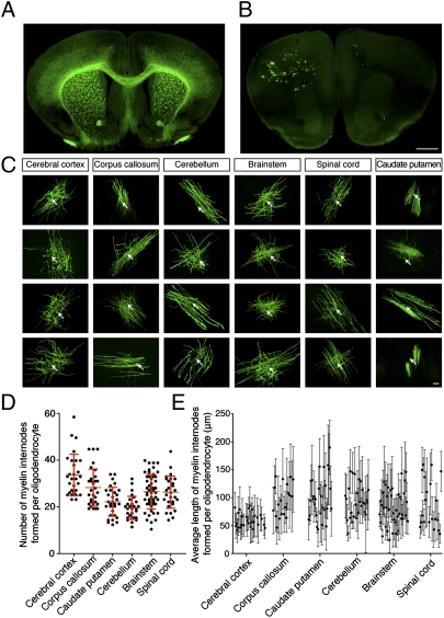
A requisite component of nervous system development is the achievement of cellular recognition and spatial segregation through competition-based refinement mechanisms. Competition for available axon space by myelinating oligodendrocytes ensures that all relevant CNS axons are myelinated properly. To ascertain the nature of this competition, we generated a transgenic mouse with sparsely labeled oligodendrocytes and establish that individual oligodendrocytes occupying similar axon tracts can greatly vary the number and lengths of their myelin internodes. Here we show that intercellular interactions between competing oligodendroglia influence the number and length of myelin internodes, referred to as myelinogenic potential, and identify the amino-terminal region of Nogo-A, expressed by oligodendroglia, as necessary and sufficient to inhibit this process. Exuberant and expansive myelination/remyelination is detected in the absence of Nogo during development and after demyelination, suggesting that spatial segregation and myelin extent is limited by microenvironmental inhibition. We demonstrate a unique physiological role for Nogo-A in the precise myelination of the developing CNS. Maximizing the myelinogenic potential of oligodendrocytes may offer an effective strategy for repair in future therapies for demyelination.


Conflict of interest statement

The authors declare no conflict of interest.

Figures

Fig. 1.
Fig. 1.
Oligodendrocytes exhibit striking diversity in the number and length of myelin internodes in vivo and in vitro. Brain section from (A) 2′,3′-cyclic nucleotide 3′-phosphodiesterase (CNP)-GFP transgenic mouse where all oligodendrocytes are fluorescently labeled. (B) Transgenic mouse expressing maGFP in <1% of oligodendrocytes. (Scale bar: 1 mm.) (C) Individual oligodendrocytes from different CNS regions of the sparsely labeled mouse. Arrows indicate cell bodies. (Scale bar: 20 μm.) (D and E) Comparison of myelinogenic potential between CNS regions. (D) Individual oligodendrocytes from indicated regions form a varied number of myelin internodes. Error bars represent SD, and the middle line represents the average number of myelin internodes formed per oligodendrocyte for each indicated region. (E) Average length of myelin internodes formed by individual oligodendrocytes. Error bars represent SD of myelin internode lengths for each individual oligodendrocyte, and demonstrate the variation of length from single cells.
Fig. 2.
Fig. 2.
Membrane-bound inhibitory cues expressed by oligodendroglia decrease the number of myelin internodes formed per oligodendrocyte. (A and B) Robust myelination is achieved as indicated by MBP in red (Right) in cocultures seeded with a high density of OPCs (A Left) and a low density of OPCs (B Left) where the majority of OPCs are replaced with polystyrene beads. OPCs are visualized by immunostaining for PDGF receptor-α (PDGFRα) axons with neurofilament (NF). Nuclei are labeled with DAPI. (Scale bar: 50 μm.) (CE) High-magnification images of an individual oligodendrocyte from high-density coculture (C), low-density coculture with polystyrene beads (D), and low-density coculture with OPC membrane-coated beads (E). (Scale bar: 20 μm.) (F) Only beads coated with OPC/oligodendrocyte membranes significantly decrease the number of internodes formed per cell in a manner comparable to high-density cocultures (P < 0.001, Tukey post hoc comparison after one-way ANOVA). All error bars represent SD unless otherwise stated.
Fig. 3.
Fig. 3.
Nogo-A is necessary and sufficient for reducing the number of myelin internodes formed per oligodendrocyte in vitro. (A) Expression of myelin-based inhibitors of axon regeneration are analyzed by Western blot in oligodendroglia, astrocytes, COS, and 3T3 cells. PDGFRα is a marker for OPCs, and MBP and MAG are markers of mature oligodendrocytes. β-actin serves as a loading control. (B) Fc-fusion candidates conjugated to Protein A-coated polystyrene beads are presented locally to oligodendroglia. (Scale bar: 50 μm.) (C) Only the amino terminal of Nogo-A (Nogo-A/Fc) significantly decreases the number of myelin internodes formed per oligodendrocyte across all conditions (P < 0.001, Tukey post hoc comparison after one-way ANOVA). (D) Knockdown of Nogo-A expression in OPC membranes before adsorption onto polystyrene beads leads to an increase in the number of myelin internodes formed per cell. Polystyrene beads are coated with membranes extracted from OPCs nucleofected with control siRNA or siRNA targeting Nogo-A. (Scale bar: 20 μm.) (E) OPC membranes treated with siRNA targeting Nogo-A or extracted from Nogo KO mice (with all isoforms of Nogo deleted) significantly rescue the number of myelin internodes formed compared with their respective controls (P < 0.0001, Student t test).
Fig. 4.
Fig. 4.
Deletion of Nogo in vivo results in enhanced myelinogenic potential and a spatial expansion of myelin in the cerebral cortex. (A and B) Deletion of Nogo in vivo results in an increase in the number of myelin internodes formed per oligodendrocyte. (A) Representative oligodendrocytes from the cerebral cortex of WT and Nogo KO. (Scale bar: 50 μm.) (B) There is a significant increase in the average number of myelin internodes made by cortical oligodendrocytes from Nogo KO compared with WT (P < 0.03, Student t test). (C Upper) Coronal views of WT and Nogo KO brains at P30. Boxes indicate locations for magnified views shown in Lower panels. (Scale bar: 1 mm.) (Lower) Exuberant myelin is seen in the upper cortical layers for the Nogo KO compared with WT. (Scale bar: 200 μm.) (D) Nogo KO mice have significantly more myelin internodes per mm2 in cortical layers 1–3 compared with WT (P < 0.0001, Student t test). (E) Magnified coronal views of the cerebral cortex at P30. OPCs are identified with PDGFRα in red and oligodendrocyte cell bodies with an antibody against Adenomatosis polyposis coli (CC1) in white. (Scale bar: 50 μm.) (F) There are significantly fewer CC1+ oligodendrocytes (P < 0.002, Student t test) and more OPCs (P < 0.007, Student t test) in the Nogo KO.
Fig. 5.
Fig. 5.
Myelinogenic potential and remyelination after lysolecithin-induced demyelination are significantly enhanced in the spinal cord of Nogo KO. (A) Representative oligodendrocytes from the white and gray matter of the spinal cord in WT and Nogo KO. (Scale bar: 50 μm.) (B) Quantification of the average number of myelin internodes made by individual oligodendrocytes from the gray and white matter of Nogo KO and WT (P < 0.0001, Student t test). (C and D Left) Light micrographs showing the dorsal funiculus of the spinal cord for WT (C) and Nogo KO (D) at 14 dpl. The lysolecithin-induced lesion sites are outlined in red. (Right) Electron micrographs of the lesion sites. Unmyelinated axons are marked with red asterisks. (Scale bar: 5 μm.) (EG) Quantification of myelin sheath thickness and myelinated axons in WT (E) and Nogo KO (F) mice by G-ratio analysis. Scatterplots display G ratios of individual axons as a function of axonal diameter. (G) Extensive remyelination is detected in lesions induced in Nogo KO (black diamonds and line) compared with WT (gray circles and line), as evident by more axons with lower G-ratio values. A G-ratio value of 1.0 represents unmyelinated axons. (HJ) Quantification of proteolipid protein (PLP) mRNA+ oligodendrocytes in lesions sites at 14 dpl for Nogo KO (H) compared with WT (I). (Scale bar: 20 μm.) (J) There is no statistically significant difference between Nogo KO and WT.

Comment in

References

    1. Baumann N, Pham-Dinh D. Biology of oligodendrocyte and myelin in the mammalian central nervous system. Physiol Rev. 2001;81:871–927. - PubMed
    1. Nave KA, Trapp BD. Axon-glial signaling and the glial support of axon function. Annu Rev Neurosci. 2008;31:535–561. - PubMed
    1. Li H, He Y, Richardson WD, Casaccia P. Two-tier transcriptional control of oligodendrocyte differentiation. Curr Opin Neurobiol. 2009;19:479–485. - PMC - PubMed
    1. Fancy SP, et al. Dysregulation of the Wnt pathway inhibits timely myelination and remyelination in the mammalian CNS. Genes Dev. 2009;23:1571–1585. - PMC - PubMed
    1. Chen Y, et al. The oligodendrocyte-specific G protein-coupled receptor GPR17 is a cell-intrinsic timer of myelination. Nat Neurosci. 2009;12:1398–1406. - PMC - PubMed

Publication types

MeSH terms