. 2011 Dec 27;108(52):21105-10.
doi: 10.1073/pnas.1111919109. Epub 2011 Dec 12.

Human monocytes are severely impaired in base and DNA double-strand break repair that renders them vulnerable to oxidative stress

Affiliations

Human monocytes are severely impaired in base and DNA double-strand break repair that renders them vulnerable to oxidative stress

Martina Bauer et al. Proc Natl Acad Sci U S A. .

Abstract

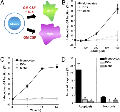
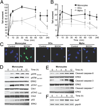
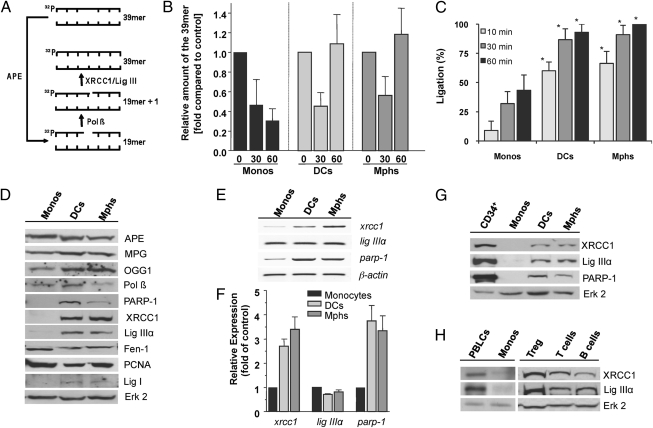
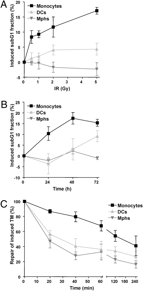
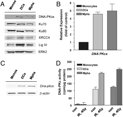
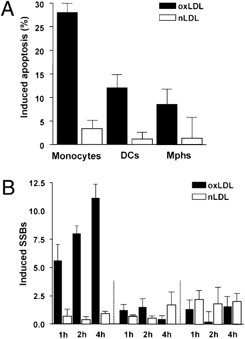
Monocytes are key players in the immune system. Crossing the blood barrier, they infiltrate tissues and differentiate into (i) macrophages that fight off pathogens and (ii) dendritic cells (DCs) that activate the immune response. A hallmark of monocyte/macrophage activation is the generation of reactive oxygen species (ROS) as a defense against invading microorganisms. How monocytes, macrophages, and DCs in particular respond to ROS is largely unknown. Here we studied the sensitivity of primary human monocytes isolated from peripheral blood and compared them with macrophages and DCs derived from them by cytokine maturation following DNA damage induced by ROS. We show that monocytes are hypersensitive to ROS, undergoing excessive apoptosis. These cells exhibited a high yield of ROS-induced DNA single- and double-strand breaks and activation of the ATR-Chk1-ATM-Chk2-p53 pathway that led to Fas and caspase-8, -3, and -7 activation, whereas macrophages and DCs derived from them were protected. Monocytes are also hypersensitive to ionizing radiation and oxidized low-density lipoprotein. The remarkable sensitivity of monocytes to oxidative stress is caused by a lack of expression of the DNA repair proteins XRCC1, ligase IIIα, poly(ADP-ribose) polymerase-1, and catalytic subunit of DNA-dependent protein kinase (DNA-PK(cs)), causing a severe DNA repair defect that impacts base excision repair and double-strand break repair by nonhomologous end-joining. During maturation of monocytes into macrophages and DCs triggered by the cytokines GM-CSF and IL-4, these proteins become up-regulated, making macrophages and DCs repair-competent and ROS-resistant. We propose that impaired DNA repair in monocytes plays a role in the regulation of the monocyte/macrophage/DC system following ROS exposure.


Conflict of interest statement

The authors declare no conflict of interest.

Figures

Fig. 1.
Fig. 1.
Death of monocytes, DCs, and macrophages following treatment with oxidizing agents. (A) Maturation of human monocytes into macrophages (Mphs) and DCs. (B) Dose dependence of cell death of monocytes, DCs, and macrophages following treatment with BOOH for 24 h. (C) Time dependence of the apoptotic response after treatment with 400 μM BOOH. Apoptosis was determined by quantifying the sub-G1 fraction by flow cytometry. (D) Frequency of induced apoptosis and necrosis of monocytes, DCs, and macrophages 24 h after treatment with 400 μM BOOH (determined by annexin V/PI double staining). *P < 0.001 for comparison of monocytes and DCs and macrophages (t test). All data are the mean of at least three independent experiments ± SD.
Fig. 2.
Fig. 2.
DNA breaks, DDR, and apoptosis in human monocytes. (A) Induced DNA SSBs following 400 μM BOOH, as determined by the alkaline comet assay. (B) Induced DSBs following 400 μM BOOH, as determined by the neutral comet assay. The level of DNA breakage was expressed by the tail moment (32). Data are the mean of at least three independent experiments ± SD. (C) γH2AX pan-staining in monocytes, DCs and macrophages (Mphs) after 1 h treatment with 400 μM BOOH. Blue, ToPro3 nuclear staining; green, γH2AX staining. (D) Western blot analysis of ATR, ATM, Chk1, and Chk2 phosphorylation and p53 stabilization in cell extracts of monocytes at different times after treatment with 400 μM BOOH. ERK2 served as loading control. (E) Time dependence of cleaved initiator caspase-8 and executor caspases 3 and 7 in cell extracts of monocytes following treatment with 400 μM BOOH. (F) Expression of fasR in monocytes, determined by semiquantitative RT-PCR. gapdh served as internal control.
Fig. 3.
Fig. 3.
BER and expression of repair proteins in monocytes, DCs, and macrophages (Mphs). (A) In vitro repair reactions were performed by using whole-cell extracts and a 32P-labeled oligonucleotide containing a single AP site. Schematic outline of the repair reaction. The 39-mer fragment (full length) represents molecules that contain the lesion or are fully repaired. (B) Relative amount of the 39-mer at increasing time points (0, 30, and 60 min). Data of three independent experiments are pooled. (C) DNA ligation assay. Relative amount of the 39-mer ligation product at increasing times of incubation (10, 30, 60 min). It was set to 100% in macrophages. Data of three independent experiments are pooled (*P < 0.001 comparing monocytes with DCs and macrophages). (D) Protein expression of APE, methylpurine-DNA glycosylase (MPG), OGG1, Pol β, Parp-1, XRCC1, Lig IIIα, Fen-1, proliferating cell nuclear antigen (PCNA), and Lig I in cell extracts of monocytes, DCs, and macrophages. Erk2 represents control. (E) mRNA level of xrcc1, lig IIIα, and parp-1 mRNA in monocytes, DCs, and macrophages, detected by semiquantitative RT-PCR. β-actin was used as internal control. (F) Expression of xrcc1, lig IIIα, and parp-1 in monocytes, DCs, and macrophages determined by quantitative RT-PCR. (G) XRCC1, Lig IIIα, and PARP-1 protein in CD34+ hematopoietic progenitor cells in comparison with monocytes, DCs, and macrophages. (H) Expression of XRCC1 and Lig IIIα in PBLCs, regulatory T cells, T cells, and B cells compared with monocytes.
Fig. 4.
Fig. 4.
Sensitivity of monocytes, DCs and macrophages to IR. (A) Apoptotic response of cells 24 h after irradiation with different doses of γ-rays. (B) Time dependence of the apoptotic response of monocytes, DCs, and macrophages after irradiation with 5 Gy. Apoptosis was determined by flow cytometry (sub-G1 fraction). (C) Kinetics of repair of induced DSBs, determined by the neutral comet assay [tail moment (TM)] after irradiation with 5 Gy. Data are the mean of at least three independent experiments ± SD.
Fig. 5.
Fig. 5.
Expression of NHEJ proteins in monocytes, DCs, and macrophages (Mphs). (A) Western blot analysis of DNA-PKcs, Ku70, Ku80, XRCC4, and Lig IV. ERK2 served as loading control. (B) Determination of mRNA expression of DNA-PKcs by quantitative PCR, demonstrating up-regulation of the mRNA level during maturation of monocytes into DCs and macrophages. (C) DNA-PKcs mRNA level determined by semiquantitative RT-PCR to demonstrate the specificity of the reaction. (D) DNA-PK activity in cell extracts of monocytes, DCs, and macrophages not treated (con) or treated with IR (4 Gy). Data are the mean of at least three independent determinations.
Fig. 6.
Fig. 6.
Genotoxic response of monocytes, DCs, and macrophages following oxLDL and cytokine production following stimulation with LPS. (A) Frequency of induced apoptosis 16 h after treatment with 200 μg/mL oxLDL and nLDL, determined by annexin V/PI double staining, in monocytes, DCs, and macrophages. (B) Formation of induced SSBs (alkaline comet assay) following treatment of cells with 200 μg/mL oxLDL and nLDL.

References

    1. Hume DA. The mononuclear phagocyte system. Curr Opin Immunol. 2006;18:49–53. - PubMed
    1. Ueno H, Schmitt N, Palucka AK, Banchereau J. Dendritic cells and humoral immunity in humans. Immunol Cell Biol. 2010;88:376–380. - PMC - PubMed
    1. Enk AH, Jonuleit H, Saloga J, Knop J. Dendritic cells as mediators of tumor-induced tolerance in metastatic melanoma. Int J Cancer. 1997;73:309–316. - PubMed
    1. Durackova Z. Some current insights into oxidative stress. Physiol Res. 2010;59:459–469. - PubMed
    1. Fialkow L, Wang Y, Downey GP. Reactive oxygen and nitrogen species as signaling molecules regulating neutrophil function. Free Radic Biol Med. 2007;42:153–164. - PubMed

Publication types

MeSH terms