. 2011 Dec 27;108(52):21182-7.
doi: 10.1073/pnas.1118450109. Epub 2011 Dec 12.

Antigen-specific CD4 T-cell help rescues exhausted CD8 T cells during chronic viral infection

Affiliations

Antigen-specific CD4 T-cell help rescues exhausted CD8 T cells during chronic viral infection

Rachael D Aubert et al. Proc Natl Acad Sci U S A. .

Abstract

CD4 T cells play a critical role in regulating CD8 T-cell responses during chronic viral infection. Several studies in animal models and humans have shown that the absence of CD4 T-cell help results in severe dysfunction of virus-specific CD8 T cells. However, whether function can be restored in already exhausted CD8 T cells by providing CD4 T-cell help at a later time remains unexplored. In this study, we used a mouse model of chronic lymphocytic choriomeningitis virus (LCMV) infection to address this question. Adoptive transfer of LCMV-specific CD4 T cells into chronically infected mice restored proliferation and cytokine production by exhausted virus-specific CD8 T cells and reduced viral burden. Although the transferred CD4 T cells were able to enhance function in exhausted CD8 T cells, these CD4 T cells expressed high levels of the programmed cell death (PD)-1 inhibitory receptor. Blockade of the PD-1 pathway increased the ability of transferred LCMV-specific CD4 T cells to produce effector cytokines, improved rescue of exhausted CD8 T cells, and resulted in a striking reduction in viral load. These results suggest that CD4 T-cell immunotherapy alone or in conjunction with blockade of inhibitory receptors may be a promising approach for treating CD8 T-cell dysfunction in chronic infections and cancer.


Conflict of interest statement

Conflict of interest statement: R.A., A.H.S., and G.J.F. hold patents for the PD-1 inhibitory pathway.

Figures

Fig. 1.
Fig. 1.
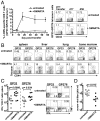
LCMV-specific transgenic CD4 T cells proliferate and persist long term after transfer into chronically infected hosts. LCMV-specific CD4 T cells were isolated from naïve Thy1.1 or Ly5.1 SMARTA transgenic mice, labeled with CFSE, and transferred into congenic B6 recipients. (A) Frequency of SMARTA cells at day 2.5 posttransfer. CFSE dilution and CD44 expression for transferred cells (gated on Thy1.1). (B) SMARTA CD4 T-cell expansion in the blood, summarized from several experiments (n = 10–15 mice per time point). (C) LCMV-specific cytokine production at 4 mo posttransfer. Shown is the percentage of SMARTA CD4 T cells producing cytokine after ex vivo stimulation with the GP61-80 peptide in one representative mouse (n = 5).
Fig. 2.
Fig. 2.
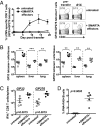
Transfer of CD4 T-cell help enhances LCMV-specific CD8 T-cell responses. (A) LCMV-specific CD8 T cells in the blood after SMARTA T-cell transfer (n = 4 untreated mice and 6–8 treated mice). Representative LCMV-specific tetramer staining (gated on CD8 T cells) is shown. (B) LCMV-specific CD8 T-cell responses in tissues at day 35 posttransfer. (C) Summarized LCMV-specific IFN-γ production by CD8 T cells in the spleen at day 7 posttransfer (n = 8 mice per group, combined from two independent experiments) and representative IFN-γ and TNF-α staining for CD8 T cells. (D) Serum viral titers as determined by plaque assay at 1 mo posttransfer. Similar results were found in several independent experiments. P values were determined by the Student t test.
Fig. 3.
Fig. 3.
Increased B-cell responses after SMARTA CD4 T-cell transfer. (A) Germinal center activity in chronically infected recipients at 1 mo after SMARTA CD4 T-cell transfer. Shown is a summary graph of the percentage of PNA+FAS+ B220+ cells in the spleen, with one representative flow plot per group. (B) At day 55 posttransfer, LCMV-specific antibody levels were measured by serum ELISA. The bottom dotted line indicates the lower limit of detection, and the top line indicates the endpoint titer in LCMV Armstrong immune mice at >60 d postinfection.
Fig. 4.
Fig. 4.
PD-1 blockade improves function but not proliferation of LCMV-specific CD4 T cells. (A) PD-1 expression on SMARTA CD4 T cells in chronically infected hosts at day 15 posttransfer. Histogram shows percent of Thy1.1 CD4 T cells expressing PD-1 compared to naive CD44low (left histogram) CD4 T cells. (B) Number of SMARTA cells in spleen (n = 3–5 mice/group). (C) Percentage of SMARTA CD4 T cells producing IFN-γ at day 15 posttransfer (n = 4 mice per group). Polyfunctionality of the SMARTA cells in chronically infected recipients after αPD-L1 blockade gated on SMARTA (Thy1.1), frequency of transferred cells producing cytokine, and mean fluorescence intensity of the IFN-γ staining.
Fig. 5.
Fig. 5.
PD-1 blockade complements CD4 T-cell therapy to enhance function in exhausted CD8 T cells and further reduce viral loads. (A) Number of LCMV-specific CD8 T cells coproducing IFN-γ and TNF-α at 2 wk posttreatment, summed from individual staining for six LCMV epitopes (GP33, GP276, NP205, NP235, GP118, and GP92). (B) Viral titers in the serum at 1 mo after CD4 T-cell transfer (LOD 50 pfu/mL). *P < 0.05; **P < 0.01; ***P < 0.001, Student t test.
Fig. 6.
Fig. 6.
Transfer of effector SMARTA CD4 T cells enhances LCMV-specific CD8 T-cell and B-cell responses in chronically infected mice. Here, 10 × 106 effector SMARTA CD4 T cells (isolated from day 7 LCMV Armstrong mice) were transferred into LCMV chronically infected recipients. (A) LCMV-specific CD8 T cells in blood (n = 5/group) and representative LCMV-specific tetramer staining (gated on CD8 T cells). (B) Total numbers of LCMV-specific CD8 T cells in tissues on day 19 posttransfer. Open symbols indicate untreated mice; closed symbols, SMARTA effector recipients. (C) Number of LCMV-specific CD8 T cells in the spleen producing IFN-γ at day 19 posttransfer. (D) Graph of average percentage and range of germinal center B cells (gated on B220+ CD19+ B cells) detected in the spleen at day 19 posttransfer. Shown is a representative experiment of two independent experiments with similar results. **P < 0.01; ***P < 0.001, Student t test. In the chronically infected mice receiving SMARTA effectors, all LCMV-specific CD8 T cells were Ly5.2, and thus were not contamination from the donor LCMV Armstrong-infected mice (Ly5.1).

References

    1. Kaech SM, Wherry EJ. Heterogeneity and cell-fate decisions in effector and memory CD8+ T cell differentiation during viral infection. Immunity. 2007;27:393–405. - PMC - PubMed
    1. Virgin HW, Wherry EJ, Ahmed R. Redefining chronic viral infection. Cell. 2009;138:30–50. - PubMed
    1. Klebanoff CA, Acquavella N, Yu Z, Restifo NP. Therapeutic cancer vaccines: Are we there yet? Immunol Rev. 2011;239:27–44. - PMC - PubMed
    1. Kim PS, Ahmed R. Features of responding T cells in cancer and chronic infection. Curr Opin Immunol. 2010;22:223–230. - PMC - PubMed
    1. Matloubian M, Concepcion RJ, Ahmed R. CD4+ T cells are required to sustain CD8+ cytotoxic T-cell responses during chronic viral infection. J Virol. 1994;68:8056–8063. - PMC - PubMed

Publication types

LinkOut - more resources