. 2012 Apr 1;302(7):F853-64.
doi: 10.1152/ajprenal.00035.2011. Epub 2011 Dec 7.

Persistent disruption of mitochondrial homeostasis after acute kidney injury

Affiliations

Persistent disruption of mitochondrial homeostasis after acute kidney injury

Jason A Funk et al. Am J Physiol Renal Physiol. .

Abstract

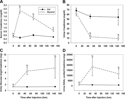
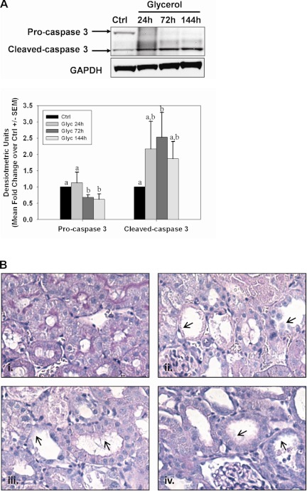
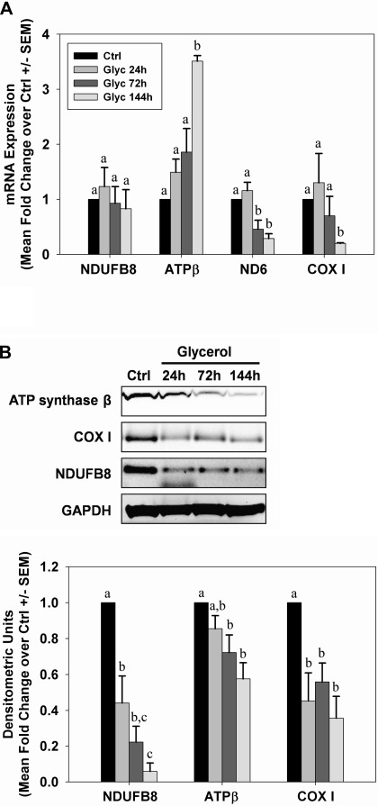
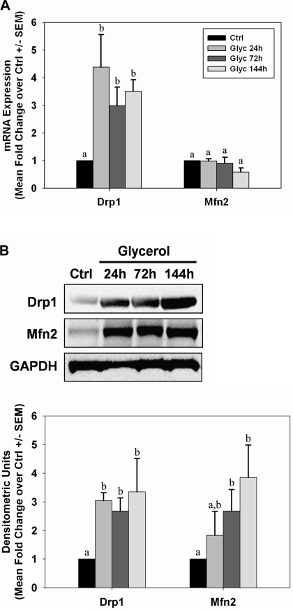
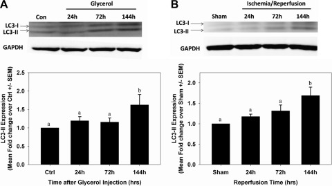
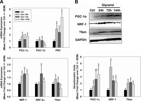
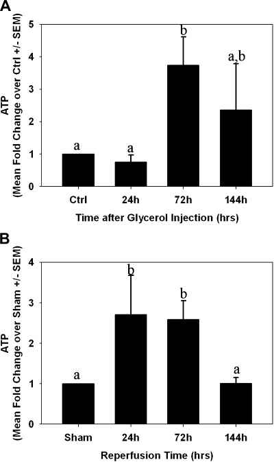
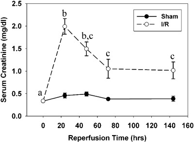
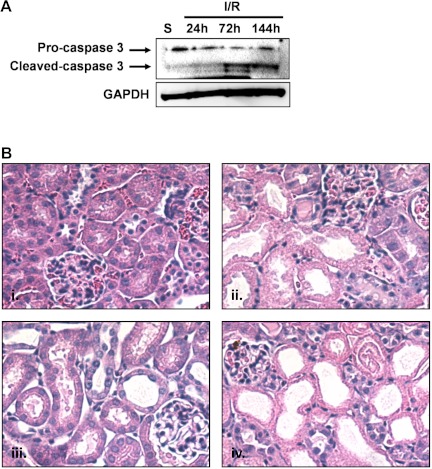
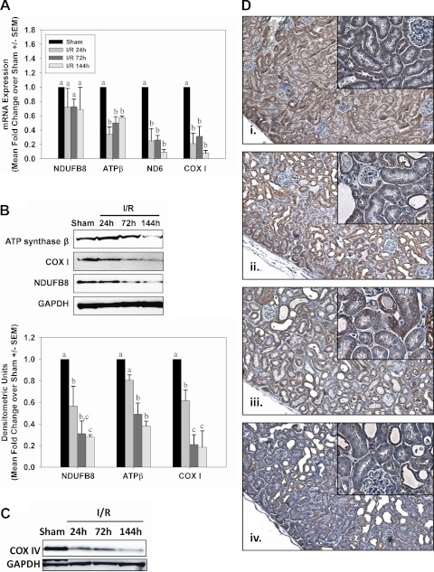
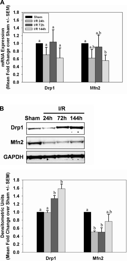
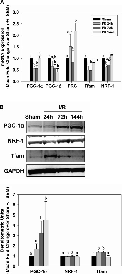
While mitochondrial dysfunction is a pathological process that occurs after acute kidney injury (AKI), the state of mitochondrial homeostasis during the injury and recovery phases of AKI remains unclear. We examined markers of mitochondrial homeostasis in two nonlethal rodent AKI models. Myoglobinuric AKI was induced by glycerol injection into rats, and mice were subjected to ischemic AKI. Animals in both models had elevated serum creatinine, indicative of renal dysfunction, 24 h after injury which partially recovered over 144 h postinjury. Markers of proximal tubule function/injury, including neutrophil gelatinase-associated lipocalin and urine glucose, did not recover during this same period. The persistent pathological state was confirmed by sustained caspase 3 cleavage and evidence of tubule dilation and brush-border damage. Respiratory proteins NDUFB8, ATP synthase β, cytochrome c oxidase subunit I (COX I), and COX IV were decreased in both injury models and did not recover by 144 h. Immunohistochemical analysis confirmed that COX IV protein was progressively lost in proximal tubules of the kidney cortex after ischemia-reperfusion (I/R). Expression of mitochondrial fission protein Drp1 was elevated after injury in both models, whereas the fusion protein Mfn2 was elevated after glycerol injury but decreased after I/R AKI. LC3-I/II expression revealed that autophagy increased in both injury models at the later time points. Markers of mitochondrial biogenesis, such as PGC-1α and PRC, were elevated in both models. These findings reveal that there is persistent disruption of mitochondrial homeostasis and sustained tubular damage after AKI, even in the presence of mitochondrial recovery signals and improved glomerular filtration.


Figures

Fig. 1.
Fig. 1.
Renal dysfunction after glycerol-induced myoglobinuria. A: serum creatinine was maximal 24 h after injection and partially recovered between 24 and 144 h after injury without returning to normal levels. B: urine creatinine was reduced 48 h after injury and remained decreased at 144 h. Urine glucose (C) and neutrophil gelatinase-associated lipocalin (NGAL; D) were elevated 48 h after glycerol injection and remained elevated at 144 h. Different superscripts above data points are significantly different from one another (P < 0.05).
Fig. 2.
Fig. 2.
Persistent tubule pathology after glycerol-induced acute kidney injury (AKI). A: activation of caspase 3 was observed by the presence of a caspase 3 cleavage fragment at 24, 72, and 144 h after glycerol injection. Bars with different superscripts are significantly different from one another (P < 0.05). B: periodic acid-Schiff (PAS) staining in control rats (i) and 24 h (ii), 72 h (iii), or 144 h (iv) after glycerol injection at ×40 magnification. Arrowheads indicate dilated tubules and brush-border damage after injury.
Fig. 3.
Fig. 3.
Sustained depletion of mitochondrial proteins after glycerol-mediated AKI. A: mRNA from control and glycerol rats was analyzed by qRT-PCR for expression of nuclear-encoded respiratory genes NDUFB8 and ATP synthase β and the mitochondrial-encoded genes ND6 and COX I at 24, 72, and 144 h after injury. B: expression of mitochondrial respiratory proteins from kidneys of control and glycerol rats was examined by immunoblot analysis. Bars with different superscripts are significantly different from one another (P < 0.05).
Fig. 4.
Fig. 4.
Alterations in mitochondrial fission and fusion proteins after glycerol-mediated AKI. mRNA (A) and protein (B) expressions from kidneys of control (Ctrl) and glycerol (Glyc) rats were analyzed by qRT-PCR and immunoblot analysis for expression of Drp1 and Mfn2 at 24, 72, and 144 h after injury. Bars with different superscripts are significantly different from one another (P < 0.05).
Fig. 5.
Fig. 5.
Induction of autophagy after AKI. LC3-I/II protein expression was measured by immunoblot analysis in control and glycerol-treated rats (A) and sham and ischemia-reperfusion (I/R) mice (B). Bars with different superscripts are significantly different from one another (P < 0.05).
Fig. 6.
Fig. 6.
Mitochondrial biogenesis after glycerol-mediated AKI. A: kidneys from control and glycerol-treated rats were analyzed for expression of genes associated with mitochondrial biogenesis by qRT-PCR. B: peroxisome proliferator-activated receptor γ co-activator 1-α (PGC-1α), nuclear respiratory factors (NRF-1), and mitochondrial transcription factor A (Tfam) protein expressions were examined in kidneys of control and glycerol-treated rats by immunoblot analysis. Bars with different superscripts are significantly different from one another (P < 0.05).
Fig. 7.
Fig. 7.
ATP levels after AKI. ATP was measured in flash-frozen kidney cortex from control/sham animals and at 24, 72, and 144 h after glycerol (A)- or I/R (B)-induced AKI. Bars with different superscripts are significantly different from one another (P < 0.05).
Fig. 8.
Fig. 8.
Kidney dysfunction after I/R injury. Serum creatinine levels were significantly elevated 24 h after reperfusion, and then slowly decreased between 24 and 144 h without returning to normal levels. Different superscripts above data points are significantly different from one another (P < 0.05).
Fig. 9.
Fig. 9.
Persistent tubule pathology after I/R AKI. A: activation of caspase 3 was observed by the presence of a caspase 3 cleavage fragment at 72 and 144 h after reperfusion. B: hematoxylin and eosin (H&E) staining in sham mice (i) and 24 h (ii), 72 h (iii), or 144 h (iv) after I/R at ×40 magnification.
Fig. 10.
Fig. 10.
Sustained depletion of mitochondrial proteins after I/R AKI. A: mRNA from sham and I/R mice was analyzed by qRT-PCR for expression of nuclear-encoded respiratory genes NDUFB8 and ATP synthase β and the mitochondrial-encoded genes ND6 and COX I at 24, 72, and 144 h after injury. B: expression of mitochondrial respiratory proteins from kidneys of sham and I/R mice was examined by immunoblot analysis. Bars with different superscripts are significantly different from one another (P < 0.05). C: immunoblot analysis confirmed reduced COX IV protein expression in kidney cortex from mice 24, 72, and 144 h after I/R. D: COX IV immunohistochemistry (brown stain) in sham mice (i) or 24 h (ii), 72 h (iii), or 144 h (iv) after reperfusion in I/R mice, with hematoxylin counterstain. Low-magnification images were captured at ×10 and higher-magnification insets were captured at ×40.
Fig. 11.
Fig. 11.
Alterations in mitochondrial fission and fusion proteins after I/R AKI. mRNA (A) and protein (B) expressions from kidneys of sham and I/R mice were analyzed by qRT-PCR and immunoblot analysis of Drp1 and Mfn2 at 24, 72, and 144 h after injury. Bars with different superscripts are significantly different from one another (P < 0.05).
Fig. 12.
Fig. 12.
Mitochondrial biogenesis after I/R AKI. A: kidneys from sham and I/R mice were analyzed for mRNA expression of genes associated with mitochondrial biogenesis by qRT-PCR. B: PGC-1α, NRF-1, and Tfam protein expressions were examined by immunoblot analysis in kidneys from sham and I/R mice. Bars with different superscripts are significantly different from one another (P < 0.05).

Similar articles

Cited by

References

    1. Bonventre JV. Mechanisms of ischemic acute renal failure. Kidney Int 43: 1160–1178, 1993 - PubMed
    1. Bonventre JV, Weinberg JM. Recent advances in the pathophysiology of ischemic acute renal failure. J Am Soc Nephrol 14: 2199–2210, 2003 - PubMed
    1. Bosch X, Poch E, Grau JM. Rhabdomyolysis and acute kidney injury. N Engl J Med 361: 62–72, 2009 - PubMed
    1. Brooks C, Wei Q, Cho SG, Dong Z. Regulation of mitochondrial dynamics in acute kidney injury in cell culture and rodent models. J Clin Invest 119: 1275–1285, 2009 - PMC - PubMed
    1. Cameron JS. Allergic interstitial nephritis: clinical features and pathogenesis. Q J Med 66: 97–115, 1988 - PubMed

Publication types