Review
. 2012 Jun;11(2):457-87.
doi: 10.1007/s12311-011-0331-9.

Consensus paper: roles of the cerebellum in motor control--the diversity of ideas on cerebellar involvement in movement

Affiliations
Review

Consensus paper: roles of the cerebellum in motor control--the diversity of ideas on cerebellar involvement in movement

Mario Manto et al. Cerebellum. 2012 Jun.

Abstract

Considerable progress has been made in developing models of cerebellar function in sensorimotor control, as well as in identifying key problems that are the focus of current investigation. In this consensus paper, we discuss the literature on the role of the cerebellar circuitry in motor control, bringing together a range of different viewpoints. The following topics are covered: oculomotor control, classical conditioning (evidence in animals and in humans), cerebellar control of motor speech, control of grip forces, control of voluntary limb movements, timing, sensorimotor synchronization, control of corticomotor excitability, control of movement-related sensory data acquisition, cerebro-cerebellar interaction in visuokinesthetic perception of hand movement, functional neuroimaging studies, and magnetoencephalographic mapping of cortico-cerebellar dynamics. While the field has yet to reach a consensus on the precise role played by the cerebellum in movement control, the literature has witnessed the emergence of broad proposals that address cerebellar function at multiple levels of analysis. This paper highlights the diversity of current opinion, providing a framework for debate and discussion on the role of this quintessential vertebrate structure.


Conflict of interest statement

Conflict of Interest The authors declare that they have no conflict of interest.

Figures

Fig. 1
Fig. 1
Mean percentage of responses collected in rabbits during classical conditioning of the nictitating membrane response. The conditioned stimulus (CS) consisted of an 800-Hz, 72-dB tone lasting for 600 ms. The unconditioned stimulus consisted of a 100-ms air puff directed at the right cornea. Nictitating membrane responses were recorded with the help of a potentiometer attached to the ipsilateral nictitating membrane. Experimental groups were as follows: the CS–UCS group received paired CS–UCS presentations. CA-A and UCS-A groups received sole presentations of CS or UCS stimuli, respectively. CS-R and UCS-R groups received unpaired presentations of CS and USC stimuli. Figure taken with permission from [33]
Fig. 2
Fig. 2
Effects of muscimol injection in, and microstimulation of, the posterior interpositus nucleus on the percentage and amplitude of conditioned eyelid responses (CRs) collected from alert behaving cats. a Diagram illustrating the experimental design. Animals were implanted with electromyographic recording electrodes in the orbicularis oculi muscle (O.O. EMG) and with a chronic guide cannula in the posterior interpositus nucleus (PIN) allowing neuronal recording (Rec), microstimulation (St), and microinjection (Inj). Animals were also implanted with stimulating electrodes in selected brain sites for antidromic identification of recorded facial motoneurons and posterior interpositus neurons [46, 50]. Delayed eyeblink conditioning was achieved by the paired presentation of a 370-ms tone used as a conditioned stimulus (CS), followed 270 ms from its start by a 100-ms air puff as an unconditioned stimulus (US). b Representative examples of CRs evoked by the sole CS presentation, collected from the fourth, fifth, and seventh conditioning sessions. Muscimol (a GABAA agonist, 1.25 μg/kg) was injected 20 min before the fifth session. The double-arrowed line (a) indicates CR amplitude. c Quantitative analysis of data collected from three animals (mean ± SEM). Muscimol was injected before the fifth and sixth sessions. Note that, according to the selected CR criterion [dashed blue line in b], the expected percentage of CRs (blue circles and dotted line) was not modified by muscimol, but the amplitude of the evoked CRs (red circles and dashed line) was significantly decreased (***p<0.001; ANOVA). d Representative examples of CRs evoked by single CS presentations without (fourth and seventh sessions) and with (fifth session) microstimulation (20 Hz for 1 s; pulses of 50 μs and 50 μA) of the posterior interpositus nucleus. e Quantitative analysis of data collected from three animals (mean ± SEM). Microstimulation was applied during the fifth and sixth sessions in trials in which the CS was presented alone. Note that, according to the selected CR criterion, the expected percentage of CRs (blue circles and dotted line) was not modified by the microstimulation, but the amplitude of the evoked CRs (green circles and dashed line) was significantly increased (**p< 0.01; ANOVA). Data collected from [46]. Figure reproduced with permission from [39]
Fig. 3
Fig. 3
Schematic representation of the reinforcing–modulating role of cerebellar interpositus neurons (IP n) during the acquisition of an associative learning task such as the classical eyeblink conditioning. This representation is based on data published elsewhere [23]. The experimental design is illustrated in Fig. 2. Neuronal inputs (green set of premotor nuclei) arriving at the orbicularis oculi motoneurons (OO MNs) and carrying eyeblink conditioned signals p(t) need the reinforcing–modulating role of cerebellar nuclei signals m(t). In order to be efficient, IP neuronal signals need to go through a learning process in order to become 180° out-of-phase with OO MN firing. Thus, IP neuronal activities (following a relay in the red nucleus) reach OO MNs right at the moment of maximum motoneuronal hyperpolarization [34], and IP neurons facilitate a quick repolarization of OO MNs, reinforcing their tonic firing during the performance of those classically conditioned eyelid responses. Abbreviation: VIIn facial nucleus. Figure reproduced with permission from [50]
Fig. 4
Fig. 4
a–c Acquisition of conditioned eyeblink responses across 3 days in patients with pure cerebellar degeneration compared to healthy controls. Mean percentage CR incidence and standard errors (SE) in paired trials (CS = tone; US = air-puff) are shown per block of ten trials and per session of 100 paired trials (total = mean total percentage CR incidence). In the group of patients, CR incidences were significantly reduced. No clear increase could be observed across the 3 days (adapted from [60])
Fig. 5
Fig. 5
Average data of grip force, load force, and acceleration obtained from vertical movements performed by three healthy subjects (a) (female, right-handed, aged 59, 63, and 65 years) and subject H. K. with cerebellar agenesis (b). Subjects moved a handheld instrumented object upward and downward. The handheld object incorporates a grip force sensor and three linear accelerations sensors registering acceleration in three dimensions including gravity. In healthy subjects, grip force starts to rise early in upward and late in downward movements (arrows). Grip and load force profiles change in parallel and peaks in load force coincide with peaks in grip force suggesting predictive force planning, regardless of movement direction. In H.K., the grip force profile does not exactly match the profile in load. Grip force starts to rise at movement onset, regardless of whether the movement is directed upward or downward (arrows). Peak grip force lags behind peak load force for upward movements, but precedes peak load force for downward movements. These findings indicate that H.K. was unable to plan and process the grip force output differentially to the direction-dependent loading requirements of the upcoming movement
Fig. 6
Fig. 6
Internal forward models enable a parallel modulation of grip force with the movement-induced loads when transporting a handheld object. The motor system generates a descending motor command that results in sensory feedback (reafference). A forward model of this system uses a copy of the descending motor command (efference copy) and generates an estimate of the sensory feedback likely to result from the movement (corollary discharge). The cerebellum computes an estimate of the sensory feedback. A mismatch between the predicted and actual sensory outcomes (prediction error) triggers force corrections along with an updating of the relevant internal models
Fig. 7
Fig. 7
Representative tasks associated with timing deficits in patients with cerebellar pathology. a Sensorimotor prediction task: SCA patients were highly variable in timing a button press to release a missile to intercept a moving target. b Temporal learning: the conditioned response is either abolished, takes longer to learn, or is produced without the appropriate delay. c A larger difference is required for the comparison interval. d Speech discrimination is disrupted when the phonetic contrast is based on the duration of a silent period (a from [122], c from [160], d from [140])
Fig. 8
Fig. 8
Absolute and relative timing. Participants are trained to make an action composed of two parts, an arm reach and a thumb button press. During training, the reach lasts 350 ms and the button press either precedes the onset of the reach (negative values), occurs during the reach (50–350 ms), or occurs after the reach is terminated (700 ms). At transfer, the participant is told to slow down the reaching movement. When the actions are successive, the absolute timing between the components is maintained. When the actions are coordinated (overlap), the timing of the thumb press is delayed to maintain relative timing. fMRI revealed a cerebellar response for the coordination condition compared to either component alone (adapted from [156])
Fig. 9
Fig. 9
Conditions (a) and behavioral results (b) in the experiment [245]. a Participants experienced illusory flexions of their right hands while viewing their video-recorded hand flexion (CONG) or extension motion (INCONG). Crosses on the wrist joints indicate fixation points. Open arrow indicates the direction of illusory movement, and solid arrows indicate the directions of visual hand motions. Three different velocities were used for each condition. b Filled bars represent the mean illusory angles across all participants under the CONG condition and open bars indicate those under the INCONG condition. Error bars indicate the standard errors of means across participants. *p<0.05
Fig. 10
Fig. 10
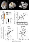
Results from the fMRI experiments [245]. a, b Right-dominant cerebral activations (a top view, b lateral view) during visuokinesthetic processing in the CONG and INCONG conditions. c Left cerebellar activations exclusively under the CONG condition. Orange and blue sections correspond to the results obtained from the right and left hands, respectively. Red section represents the area where strength of activity correlated with the subjective experience of hand movement. The horizontal plane (z=−27) is displayed. d The size of effects of left cerebellar activation (orange in c) across conditions. Bars indicate the means of contrast parameter estimates (size of effect in arbitrary units) for the left cerebellar activation (−27, −69, −30) during the CONG (orange bar), INCONG (black bar), and other control conditions (open bars; see [245] for details). Error bars represent standard errors of means across participants. e Significant correlation between the behavioral ratings (illusion scores) and the left cerebellar activity (red in c; r=0.57, df=34, p<0.001 one-tailed). The illusion scores are mean-corrected. f Relationship of activities between the right IPL (Dashed yellow circle in panel b) and the left cerebellum in a representative participant, revealed by functional connectivity analysis. The regression slopes were 0.52 and 0.29 for the CONG and INCONG conditions, respectively. The activities (x-axis for right IPL; y-axis for left cerebellum) are mean-adjusted (arbitrary units)
Fig. 11
Fig. 11
The intrinsically connected motor network at rest (from [251]). a Axial slices (Arabic numbers indicate z-coordinates). b Coronal slices passing through the cerebellum (Arabic numbers indicate y-coordinates). Abbreviations: LN lentiform nucleus (pallidum), M1 motor cortex, PMC lateral premotor cortex (clusters also include insula/claustrum), SMA supplementary motor cortex, THAL thalamus

References

    1. Baier B, Stoeter P, Dieterich M. Anatomical correlates of ocular motor deficits in cerebellar lesions. Brain. 2009;132:2114–24. - PubMed
    1. Ohki M, Kitazawa H, Hiramatsu T, Kaga K, Kitamura T, Yamada J, Nagao S. Role of primate cerebellar hemisphere in voluntary eye movement control revealed by lesion effects. J Neurophysiol. 2009;101(2):934–47. - PubMed
    1. Hiramatsu T, Ohki M, Kitazawa H, Xiong G, Kitamura T, Yamada J, Nagao S. Role of primate cerebellar lobulus petrosus of paraflocculus in smooth pursuit eye movement control revealed by chemical lesion. Neurosci Res. 2008;60(3):250–8. - PubMed
    1. Zee DS, Leigh RJ, Mathieu-Millaire F. Cerebellar control of ocular gaze stability. Ann Neurol. 1980;7:37–40. - PubMed
    1. Zee DS, Yamazaki A, Butler PH, Gücer G. Effects of ablation of flocculus and paraflocculus of eye movements in primate. J Neurophysiol. 1981;46:878–99. - PubMed

LinkOut - more resources