. 2012 May;109(5):1293-304.
doi: 10.1002/bit.24399. Epub 2011 Dec 20.

384 hanging drop arrays give excellent Z-factors and allow versatile formation of co-culture spheroids

Affiliations

384 hanging drop arrays give excellent Z-factors and allow versatile formation of co-culture spheroids

Amy Y Hsiao et al. Biotechnol Bioeng. 2012 May.

Abstract

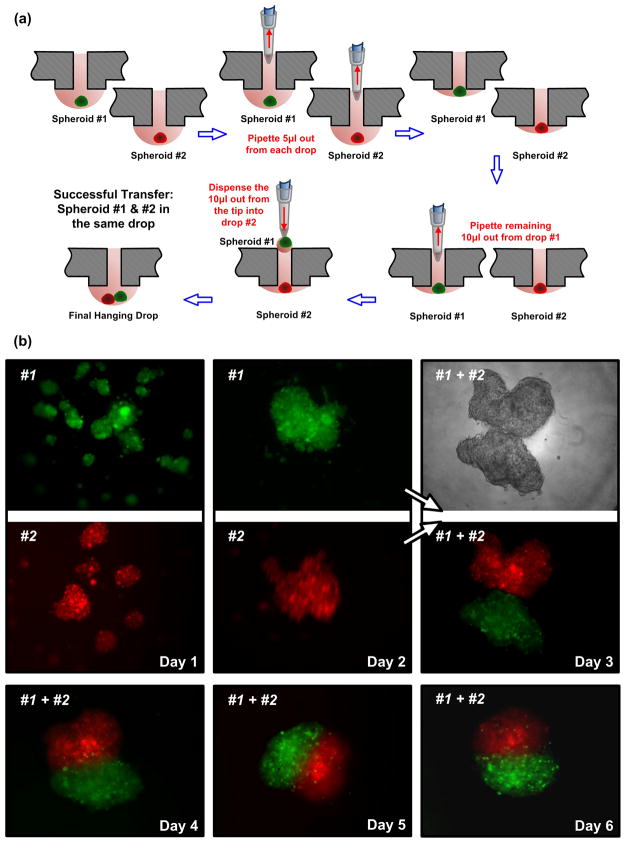
We previously reported the development of a simple, user-friendly, and versatile 384 hanging drop array plate for 3D spheroid culture and the importance of utilizing 3D cellular models in anti-cancer drug sensitivity testing. The 384 hanging drop array plate allows for high-throughput capabilities and offers significant improvements over existing 3D spheroid culture methods. To allow for practical 3D cell-based high-throughput screening and enable broader use of the plate, we characterize the robustness of the 384 hanging drop array plate in terms of assay performance and demonstrate the versatility of the plate. We find that the 384 hanging drop array plate performance is robust in fluorescence- and colorimetric-based assays through Z-factor calculations. Finally, we demonstrate different plate capabilities and applications, including: spheroid transfer and retrieval for Janus spheroid formation, sequential addition of cells for concentric layer patterning of different cell types, and culture of a wide variety of cell types.


Figures

Figure 1
Figure 1
(a) An actual image of the 384 hanging drop array plate highlighting the key features and specifications. (b) Concentration map in 384-format plate for fluorescein solution. (c) Concentration map in 384-format plate for yellow food color solution. (d) Bar graph showing the summary of Z-factors for fluorescence-based assay at various fluorescein concentrations. (e) Bar graph showing the summary of Z-factors for colorimetric-based assay at various percentages of yellow food color.
Figure 2
Figure 2
(a) Time-lapse images of mES-Oct4-GFP cell embryoid bodies cultured in the 384 hanging drop array plate at two initial seeding densities. GFP green fluorescence represents cells that still express Oct4. (b) Time-lapse images of HepG2 cell spheroids cultured in the 384 hanging drop array plate at two initial seeding densities. On Day 13, a viability staining was performed where green represents live cells and red represents dead cells. All embyroid bodies and spheroids were cultured in 15 μL hanging drops. Scale bar is 200 μm.
Figure 3
Figure 3
(a) Time-lapse images of DU145Luc prostate cancer spheroids cultured in the 384 hanging drop array plate at three initial seeding densities. On Day 8, a viability staining was performed where green represents live cells and red represents dead cells. (b) Time-lapse images of HFOB spheroids cultured in the 384 hanging drop array plate at three initial seeding densities. On Day 7, a viability staining was performed where green represents live cells and red represents dead cells. All spheroids were cultured in 15 μL hanging drops. Scale bar is 200 μm.
Figure 4
Figure 4
(a) Phase and fluorescent images of a PC-3DsRed, HUVEC, MC3T3-E1 (1:50:50 ratio) mixed co-culture spheroid. (b) Images of PC-3DsRed and MC3T3-E1 (1:100 ratio) co-culture spheroids with PC-3DsRed cells preferentially patterned in the center, exterior, or randomly distributed. (c) Time-lapse images of CellTracker Green- and Red-labeled COS7 cells patterned into concentric layers within a spheroid, where CellTracker Red-labeled COS7 cells were added to the hanging drop on Day3 and thereafter attach to the outside periphery of the existing green spheroid. All spheroids were initially cultured in 15 μL hanging drops. For the concentrically patterned spheroids in (b) and (c), 5 μL of the second cell suspension was subsequently added to the existing 15 μL droplets, bringing the final total hanging drop volume to 20 μL. Scale bar is 200 μm.
Figure 5
Figure 5
(a) Cartoon illustrating the process of spheroid transfer between hanging drops in the 384 hanging drop array plate. Initially each spheroid is cultured in a 15 μL hanging drop, and 5 μL of media is removed from each drop. All the remaining 10 μL from drop #1 (including spheroid #1) is subsequently pipetted and transferred to the remaining 10 μL of drop #2. The final drop contains both spheroid #1 and spheroid #2 in 20 μL of media. (b) Actual time-lapse images of CellTracker Green- and Red-labeled COS7 spheroids before (Day 1 and 2) and after (Day 3, 4, 5, and 6) transfer between hanging drops in the 384 hanging drop array plate. COS7 cells formed into multiple small spheroids per hanging drop on Day 1 and subsequently aggregated into single spheroid per drop by Day 2. Upon transferring the green spheroid (#1) into the hanging drop containing the red spheroid (#2), the two spheroids slowly aggregated together over the next 4 days and formed into a single Janus spheroid by Day 6.

References

    1. Baraniak PR, McDevitt TC. Scaffold-free culture of mesenchymal stem cell spheroids in suspension preserves multilineage potential. Cell Tissue Res. 2011 doi: 10.1007/s00441-011-1215-5. - DOI - PMC - PubMed
    1. Birmingham A, Selfors LM, Forster T, Wrobel D, Kennedy CJ, Shanks E, Santoyo-Lopez J, Dunican DJ, Long A, Kelleher D, Smith Q, Beijersbergen RL, Ghazal P, Shamu CE. Statistical methods for analysis of high-throughput RNA interference screens. Nat Methods. 2009;6:569–575. - PMC - PubMed
    1. Bratt-Leal AM, Kepple KL, Carpenedo RL, Cooke MT, McDevitt TC. Magnetic manipulation and spatial patterning of multi-cellular stem cell aggregates. Integr Biol. 2011 doi: 10.1039/C1IB00064K. - DOI - PMC - PubMed
    1. Friedrich J, Ebner R, Kunz-Schughart LA. Experimental anti-tumor therapy in 3-D: spheroids- old hat or new challenge. Int J Radiat Biol. 2007;83:849–871. - PubMed
    1. Friedrich J, Seidel C, Ebner R, Kunz-Schughart LA. Spheroid-based drug screen: considerations and practical approach. Nat Protoc. 2009;4:309–324. - PubMed

Publication types