. 2011 Nov;70(5):805-21.
doi: 10.1002/ana.22527.

Recovery from chronic spinal cord contusion after Nogo receptor intervention

Affiliations

Recovery from chronic spinal cord contusion after Nogo receptor intervention

Xingxing Wang et al. Ann Neurol. 2011 Nov.

Abstract

Objective: Several interventions promote axonal growth and functional recovery when initiated shortly after central nervous system injury, including blockade of myelin-derived inhibitors with soluble Nogo receptor (NgR1, RTN4R) decoy protein. We examined the efficacy of this intervention in the much more prevalent and refractory condition of chronic spinal cord injury.

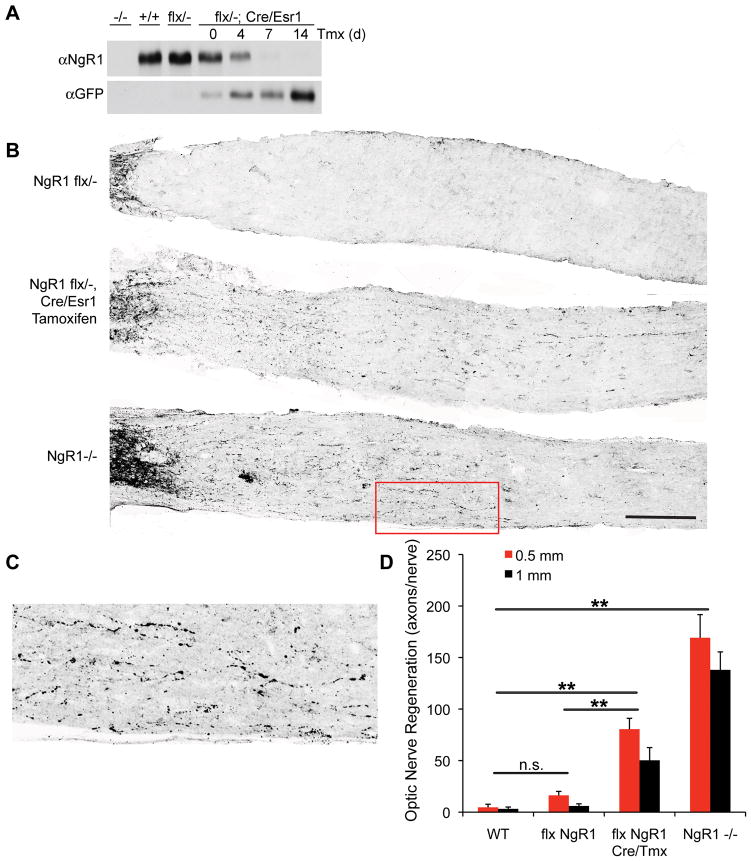
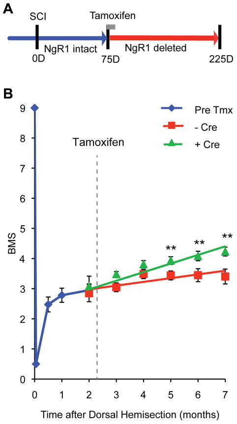
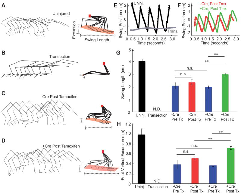
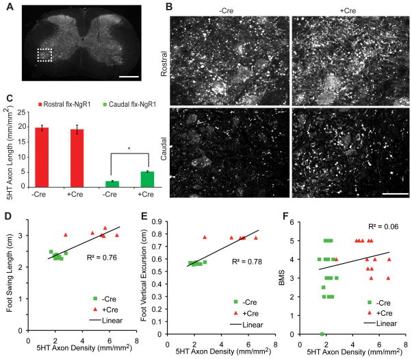
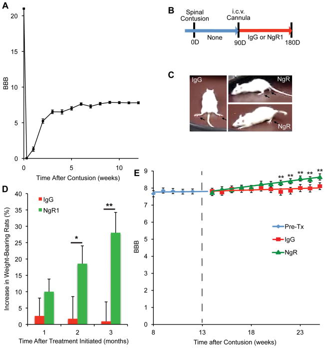
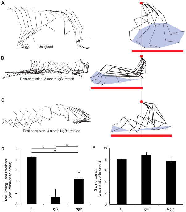
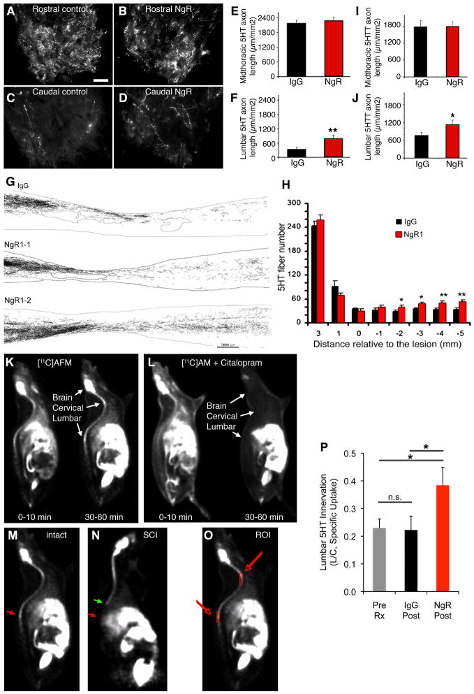
Methods: We eliminated the NgR1 pathway genetically in mice by conditional gene targeting starting 8 weeks after spinal hemisection injury and monitored locomotion in the open field and by video kinematics over the ensuing 4 months. In a separate pharmacological experiment, intrathecal NgR1 decoy protein administration was initiated 3 months after spinal cord contusion injury. Locomotion and raphespinal axon growth were assessed during 3 months of treatment between 4 and 6 months after contusion injury.

Results: Conditional deletion of NgR1 in the chronic state results in gradual improvement of motor function accompanied by increased density of raphespinal axons in the caudal spinal cord. In chronic rat spinal contusion, NgR1 decoy treatment from 4 to 6 months after injury results in 29% (10 of 35) of rats recovering weight-bearing status compared to 0% (0 of 29) of control rats (p < 0.05). Open field Basso, Beattie, and Bresnahan locomotor scores showed a significant improvement in the NgR-treated group relative to the control group (p < 0.005, repeated measures analysis of variance). An increase in raphespinal axon density caudal to the injury is detected in NgR1 decoy-treated animals by immunohistology and by positron emission tomography using a serotonin reuptake ligand.

Interpretation: Antagonizing myelin-derived inhibitors signaling with NgR1 decoy augments recovery from chronic spinal cord injury.


Figures

Figure 1
Figure 1. Recovery from Chronic SCI Deficits after NgR1 Deletion
A, Cre-mediated recombination from the pActin-Cre/Esr1 transgene abolishes expression of NgR1 following tamoxifen injection. Immunoblots of brain lysate from mice transgenic for pActin-Cre/Esr1 at several time points after tamoxifen injection to activate the Cre fusion protein with antibodies directed against either NgR1 or GFP. B, Mice of the indicated genotypes underwent optic nerve crush injury at 10–12 weeks of age, and tissue was collected 14 days later for anti-GAP-43 immunohistology of injured fibers. The flx NgR1 mice carrying the cre/Esr1 transgene were treated with tamoxifen one week prior to injury. Intact fibers close to the eye are visible at the far left, and regeneration past the lesion is detected in the bottom two panels. Scale bar, 250 μm. C, Higher magnification view of the area boxed in red of B demonstrates regenerating fibers in NgR1−/− mice. D, The number of regenerating optic nerve fibers is presented as a function of distance central to the crush site and of genotype. Data are mean ± sem for 4–8 mice per group. **, P<0.01, one-way ANOVA with pairwise post-hoc Fisher’s least-significant difference test, SPSS. The indicated comparisons are valid at both the 0.5 mm and the 1 mm distances.
Figure 2
Figure 2. Recovery of Locomotor Function in Chronic Spinal Cord Hemisection by NgR1 Deletion
A, Schematic of mouse chronic SCI experiment. All mice carried flx NgR1 alleles, but one group also carried the Cre/Esr1 transgene to allow tamoxifen-induced recombination (Cre-positive, n=23) while the other group did not (Cre-negative, n=18). 75 days after SCI, mice received tamoxifen to abolish NgR1 expression. B, The BMS score after the initiation of treatment is plotted. Recovery is greater in the Cre/Esr1 group, P=0.030 by repeated measures ANOVA for the effect of Cre after tamoxifen treatment. Data are mean ± sem. **, P<0.01, one-way ANOVA for the Cre-positive group relative to pre-tamoxifen value by Fisher’s least-significant difference test using SPSS software.
Figure 3
Figure 3. Kinematic Analysis of Hindlimb Function after NgR1 Deletion in Chronic SCI
A–D, Hindlimb kinematic analysis from individual mice with no injury (A), or a complete transection 7 days earlier (B) or dorsal hemisection 6 months earlier (C, D). The dorsal hemisection mice with flx NgR1 alleles were treated with tamoxifen 75 days after injury after injury and either carried Cre transgene (D) or not (C). Tracings of joint and limb position from one hindlimb during one gait cycle are shown for every 21st frame at the left. On the right, the foot, ankle and knees have been normalized to the hip position (red dots). The extent of foot swing is highlighted (dotted lines). E, F, The anterior-posterior position of the foot relative to the hip from one limb of each of the indicated groups as in A-D is plotted as a function of time. Gait cycles are reflected in sequential peaks. G, The length of the foot swing relative to the hip in the anterior-posterior dimension is shown for each group of mice. Mice were examined when uninjured, one week after complete spinal cord transection, or 10–35 weeks after dorsal hemisection injury. The Pre Tx groups were analyzed immediately before tamoxifen (10–11 weeks post injury), and the Post Tx groups were 4–5 months after tamoxifen (30–35 weeks post injury). Data are mean ± sem, n= 46 limbs for mice without Cre and n=36 for mice with Cre transgene. **, P<0.01, one-way ANOVA with pairwise post-hoc Fisher’s least-significant difference test, SPSS. H, The vertical excursion of the foot relative to the hip is reported for the same groups of mice as in G. Data are mean ± sem. **, P<0.01, one-way ANOVA with pairwise post-hoc analysis using SPSS software.
Figure 4
Figure 4. Raphespinal Growth Induced by NgR1 Deletion in Chronic SCI
A, Low magnification transverse section of the thoracic spinal cord indicating the ventral horn region shown at high magnification in B (dotted lines). Scale bar, 500 μm. B, Immunoreactive 5HT fibers in the ventral horn of the cervical (Rostral) and lumbar (Caudal) enlargements are shown from a mouse with or with the Cre transgene following dorsal hemisection and tamoxifen treatment at 35 weeks after injury. Scale bar, 50 μm. C, The length of 5HT immunoreactive axonal length in B. Data are mean ± sem, n = 23 mice for –Cre and n= 18 mice for +Cre group. *, P<0.01, one-way ANOVA with pairwise post-hoc analysis using SPSS software. D–F, Correlation between 5HT immunoreactive axonal length and the kinematic analysis from Fig. 3 or the BMS data from Fig. 2. Correlation coefficients from linear regression are shown.
Figure 5
Figure 5. Recovery of weight bearing after NgR1 treatment of chronically spinal cord contused rats
A, Spontaneous improvement of open field locomotion (BBB score) as a function of time after thoracic spinal cord contusion injury for all rats prior to randomization to IgG or NgR1 treatment. Mean ± sem, n = 64. B, Schematic of experiment. Two weeks after the i.c.v. cannula implantation (12 weeks post-contusion injury), rats were assigned to one of two treatment groups. The PBS minipumps were replaced with new osmotic minipumps filled with 2.25 mg AA-NgR(310)ecto-Fc (0.29 mg/kg/day) or 2.25 mg rat IgG in 2 ml PBS. The duration of treatment was 12 weeks. A new osmotic mimipump filled with same amount of AA-NgR(310)ecto-Fc or rat IgG replaced each depleted pump every 4 weeks. C, Examples of a control rat without weight support at the end of the treatment period and two of the seven AA-NgR(310)ecto-Fc treated rats that regained weight support. D, The increase in the percentage of rats showing body weight support with at least one hindlimb as a function of time during therapy is reported. P=0.022 by repeated measures ANOVA for the effect of NgR1 versus IgG treatment, and *, P<0.05, and **, P<0.01 for the indicated comparisons by one-way ANOVA. E, The course of recovery after the initiation of treatment (dotted line) is plotted as a function of time. The locomotor BBB scores from the IgG and AA-NgR(310)ecto-Fc-treated groups were indistinguishable at the initiation of treatment. Data are mean ± sem, n=29 for IgG and n=35 for NgR1 group. P=0.002 by repeated measures ANOVA for the effect of NgR1 versus IgG treatment, and **, P<0.01, one-way ANOVA for NgR1 treated values relative to pre-treatment value by post-hoc Fisher’s least-significant difference test using SPSS software.
Figure 6
Figure 6. Gait kinematics in chronic SCI rats after NgR1 treatment
A–C, Stick tracing of joint and limb position in the anterior-posterior plane (left), with forward to right, and every 16th frame shown. The limb positions after normalizing to the iliac crest position (red dot) are provided at right. The total extent of foot swing (red line) and the toe cycle (gray fill) are shown. The individual examples are of an uninjured animal (A), and of rats after treatment for 12 weeks with IgG (B) or with AA-NgR(310)ecto-Fc (C). D, The mid position of the foot swing relative to the iliac crest during the gait cycle in the different treatment groups. Data are mean ± sem for n = 12–14 limbs per condition. UI, uninjured. *, P<0.05 by one-way ANOVA with post-hoc pairwise comparison, SPSS. E, The anterior-posterior extent of foot movement relative to the iliac crest for the different treatment groups. Data are mean ± sem for n = 12–14 limbs per condition.
Figure 7
Figure 7. AA-NgR(310)ecto-Fc-induced raphespinal growth in chronic SCI
A–D, Transverse section from the spinal cord 11–15 mm rostral or caudal to the contusion site were immunostained as indicated from rats completing 3 months of i.c.v. treatment. Anti-serotonin immunohistochemistry labels 5-HT fibers in the ventral horn of the spinal cord. Note the decreased innervation caudal to the injury and the partial recovery in the AA-NgR(310)ecto-Fc treated rats. Scale bar, 25 μm. E, F, The length of 5HT-immunoreactive axon per unit area of ventral horn in the transverse plane was measured for chronic SCI rats completing 3 months treatment with the indicated proteins from micrographs as in A-D. The data are mean ± sem for n=21–25 per group, and the increase in the NgR1 treated group in the distal cord is significant; **, P<0.01, ANOVA. G, Camera lucida drawings of serotonergic fibers from one IgG-treated and two AA-NgR(310)ecto-Fc-treated animals. Each drawing is a composite assembled from a set of 10 parasagittal sections spaced at intervals of 200 μm across the spinal cord. The contusion cavities are encircled near the center of each image. Increased numbers of serotonergic fibers are observed in the caudal spinal cord in the AA-NgR(310)ecto-Fc-treated (NgR1–1, NgR1–2) animals compared with the IgG-treated animals. Scale bar is 1000 μm. H, Serotonergic (5HT) fiber number at various distances rostral and caudal to the center of the lesion from AA-NgR(310)ecto-Fc-treated (red bars) and control animals (black bars) is reported. *, P<0.05; **, P<0.01, ANOVA. For the x-axis, a positive value is rostral to the center of the lesion, and a negative value is caudal to the center of lesion. I, J, Adjacent sections were processed for anti-serotonin transporter (5HTT) immunohistochemistry and measured as for 5HT in E, F. The data are mean ± for n=21–25 per group, and the increase in the NgR1 group in the distal cord is significant, P<0.05, ANOVA, *. K, L, [11C]AFM radioactivity was visualized at the indicated times after an intravenous injection of tracer (50 ± 30 MBq, 0.12 ± 0.09 μg) with or without the pre-injection of the competitive ligand, citalopram (2 mg/kg, 15 min before [11C]AFM injection). The initial distribution of the tracer is broad with or without citalopram at 0–10 min. Specific uptake of [11C]AFM at 30–60 min in the brain, cervical spinal cord and lumbar spinal cord is blocked by citalopram. M, N, A rat with an intact spinal cord was imaged with [11C]AFM and compared to a rat with a midthoracic transection one week earlier. Uptake at 30–60 minute is illustrated. The site of the lesion is indicated by the green arrow and the reduced uptake in the lumbar cord of the injured rat is indicated by the red arrow. O, For quantitation of [11C]AFM uptake in the spinal cord, two regions of interest were selected as illustrated by the boxes on this intact rat image. P, The ratio of lumbar to cervical [11C]AFM specific uptake at 30–60 minute post-tracer injection was determined in injured rats completing 3 months of treatment with control IgG or NgR(310)ecto-Fc, as illustrated in O. The data are mean ± sem for n=21–23 per group, and the increase in the NgR1 group in the distal cord is significant; *, P<0.05, ANOVA.

References

    1. Rossignol S, Schwab M, Schwartz M, Fehlings MG. Spinal cord injury: time to move? J Neurosci. 2007;27:11782–11792. - PMC - PubMed
    1. Liu BP, Cafferty WB, Budel SO, Strittmatter SM. Extracellular regulators of axonal growth in the adult central nervous system. Philos Trans R Soc Lond B Biol Sci. 2006;361:1593–1610. - PMC - PubMed
    1. Maier IC, Schwab ME. Sprouting, regeneration and circuit formation in the injured spinal cord: factors and activity. Philos Trans R Soc Lond B Biol Sci. 2006;361:1611–1634. - PMC - PubMed
    1. Bradbury EJ, McMahon SB. Spinal cord repair strategies: why do they work? Nat Rev Neurosci. 2006;7:644–653. - PubMed
    1. Courtine G, Song B, Roy RR, et al. Recovery of supraspinal control of stepping via indirect propriospinal relay connections after spinal cord injury. Nat Med. 2008;14:69–74. - PMC - PubMed

Publication types

MeSH terms