

Altered functional magnetic resonance imaging resting-state connectivity in periaqueductal gray networks in migraine
- PMID: 22162064
- PMCID: PMC3243965
- DOI: 10.1002/ana.22537
Altered functional magnetic resonance imaging resting-state connectivity in periaqueductal gray networks in migraine
Abstract
Objective: The periaqueductal gray matter (PAG), a known modulator of somatic pain transmission, shows evidence of interictal functional and structural abnormalities in migraineurs, which may contribute to hyperexcitability along spinal and trigeminal nociceptive pathways, and lead to the migraine attack. The aim of this study was to examine functional connectivity of the PAG in migraine.
Methods: Using resting-state functional MRI, we compared functional connectivity between PAG and a subset of brain areas involved in nociceptive/somatosensory processing and pain modulation in 17 subjects with migraine, during a pain-free state, versus 17 gender- and age-matched controls. We also assessed the relation between intrinsic resting-state correlations within PAG networks and the average monthly frequency of migraine attacks, as well as allodynia.
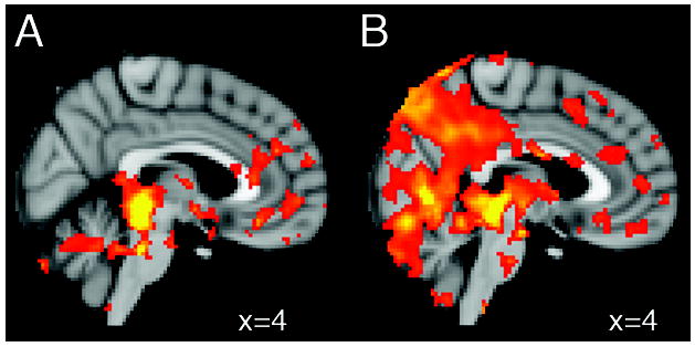
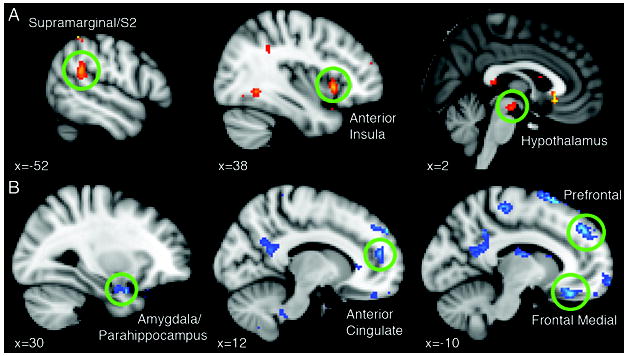
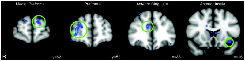
Results: Our findings show stronger connectivity between the PAG and several brain areas within nociceptive and somatosensory processing pathways in migraineurs versus controls. In addition, as the monthly frequency of migraine attacks worsens, the strength of the connectivity in some areas within these pathways increases, whereas a significant decrease in functional resting-state connectivity between the PAG and brain regions with a predominant role in pain modulation (prefrontal cortex, anterior cingulate, amygdala) can be evidenced. Finally, migraineurs with a history of allodynia exhibit significantly reduced connectivity between PAG, prefrontal regions, and anterior cingulate compared to migraineurs without allodynia.
Interpretation: These data reveal interictal dysfunctional dynamics within pain pathways in migraine manifested as an impairment of the descending pain modulatory circuits, likely leading to loss of pain inhibition, and hyperexcitability primarily in nociceptive areas.
Copyright © 2011 American Neurological Association.
Figures
Comment in
-
Pathophysiology of migraine.Headache. 2013 Feb;53(2):420-2. doi: 10.1111/head.12027. Headache. 2013. PMID: 23560278 No abstract available.
References
Publication types
MeSH terms
Grants and funding
LinkOut - more resources
Full Text Sources
Medical
