. 2011 Dec 12;195(6):1017-31.
doi: 10.1083/jcb.201105166.

Distinct intracellular motifs of Delta mediate its ubiquitylation and activation by Mindbomb1 and Neuralized

Affiliations

Distinct intracellular motifs of Delta mediate its ubiquitylation and activation by Mindbomb1 and Neuralized

Aikaterini Daskalaki et al. J Cell Biol. .

Abstract

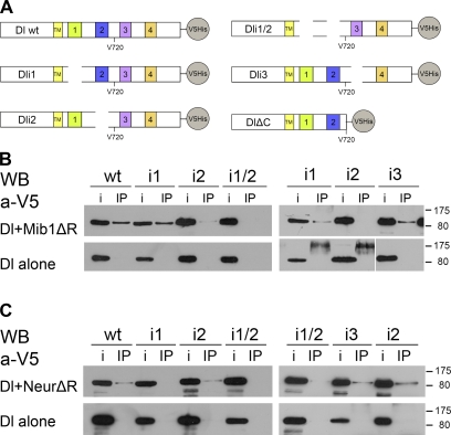
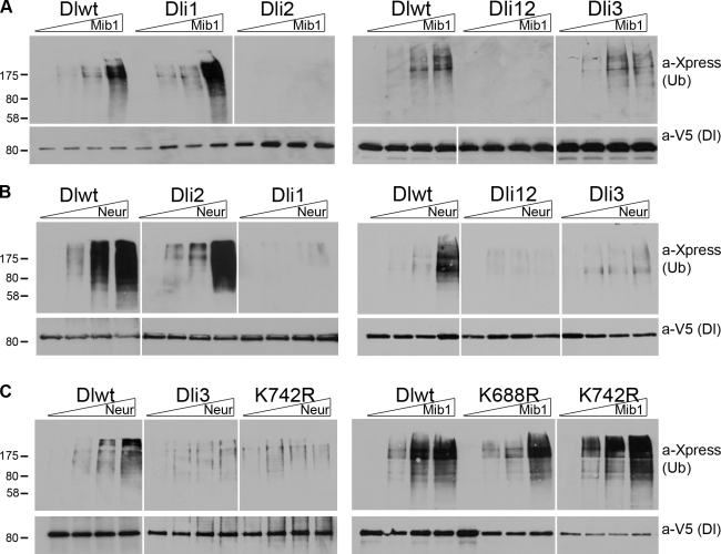
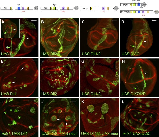
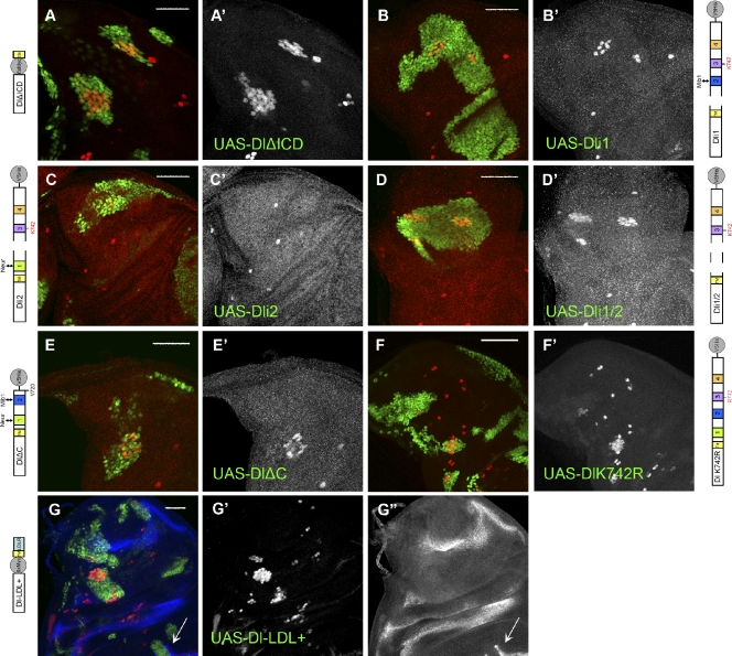
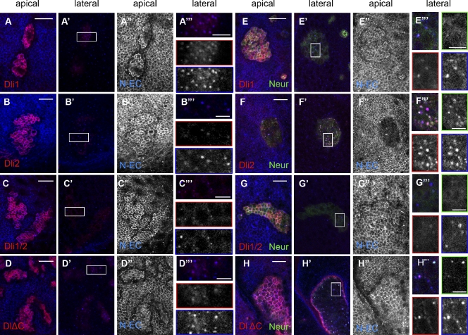
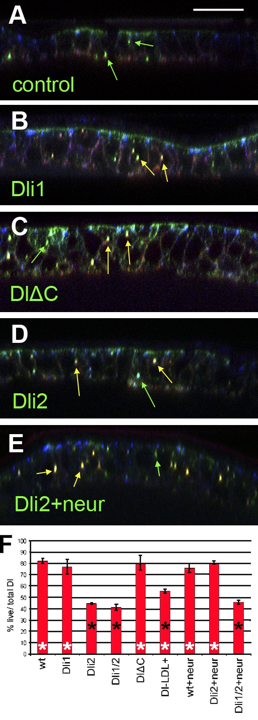
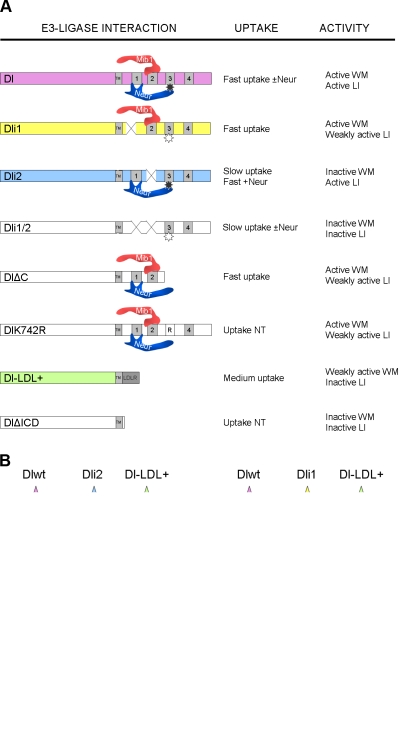
DSL proteins are transmembrane ligands of the Notch receptor. They associate with a RING (really interesting new gene) family E3 ubiquitin ligase, either Neuralized (Neur) or Mindbomb 1 (Mib1), as a prerequisite to signaling. Although Neur and Mib1 stimulate internalization of DSL ligands, it is not known how ubiquitylation contributes to signaling. We present a molecular dissection of the intracellular domain (ICD) of Drosophila melanogaster Delta (Dl), a prototype DSL protein. Using a cell-based assay, we detected ubiquitylation of Dl by both Neur and Mib1. The two enzymes use distinct docking sites and displayed different acceptor lysine preferences on the Dl ICD. We generated Dl variants that selectively perturb its interactions with Neur or Mib1 and analyzed their signaling activity in two in vivo contexts. We found an excellent correlation between the ability to undergo ubiquitylation and signaling. Therefore, ubiquitylation of the DSL ICD seems to be a necessary step in the activation of Notch.


Figures

Figure 1.
Figure 1.
Dl interactions with Neur and Mib1. (A) Schematic of Dl and its variants tested in this study. TM, transmembrane domain. ECDs and ICDs are not drawn to scale. For the exact extent of motifs 1–4, refer to Fig. S1. (B) C-terminally V5-tagged Dl variants, as indicated, were expressed alone (bottom) or with Myc-tagged Mib1ΔR (top). Immunoprecipitation (IP) lanes: extracts were immunoprecipitated with anti-Myc and detected with anti-V5. Input (i) lanes: 4% of the total extract from the same transfection was kept before immunoprecipitating the rest. The main band is full-length Dl (predicted MM of 90 kD). The bands visible in the bottom right near 175 kD are nonspecific cross-reacting bands. Marker sizes are shown on the right in kilodaltons. The white line indicates that intervening lanes have been spliced out. (C) The same Dl variants were expressed alone (bottom) or with NeurΔR (top). Extracts were immunoprecipitated with an anti-Neur antiserum and detected with anti-V5. Labels are the same as in B. WB, Western blot.
Figure 2.
Figure 2.
Ubiquitylation of Dl variants. (A) Dl-expressing constructs were cotransfected with Xpress-Ub and increasing amounts (0, 0.5, 1, and 1.5 µg) of Mib1-expressing constructs. Dl protein was isolated by affinity purification on a Ni2+ resin under denaturing conditions. Eluates of the Ni2+ column were probed with anti-Xpress to detect ubiquitylated species (top shows 1/4 of total loaded) and anti-V5 to detect total Dl as a loading control (bottom shows 1/3 of total loaded). Note that ubiquitylated species, where present, run at much higher MMs than the expected 90 kD of unmodified Dl. (B) Dl-expressing constructs were cotransfected with Xpress-Ub and increasing amounts (0, 0.5, 1, and 1.5 µg) of Neur-expressing constructs. Note weaker levels of Ub signal in Dli3. (C) Ubiquitylation assays performed as in A and B using the Dl variants and E3 ligases shown at the top. Note that Dl-K742R is weakly ubiquitylated by Neur but resembles wt Dl in its response to Mib1. MM markers are shown in kilodaltons. White lines indicate that intervening lanes have been spliced out.
Figure 3.
Figure 3.
Induction of Wg by Dl variants. Third instar wing pouches overexpressing Dl variants and stained for Wg (red) are shown. Anterior is to the left and ventral is down. (A–D and H) Dl variants as indicated are expressed with ptc-Gal4 and detected with anti-Dl. (A, inset) Disk expressing wt Dl under ptc-Gal4. Note ectopic Wg expression posterior to the Dl stripe. The narrow stripe of Dl expression sometimes seen (green arrows) comes from the overlying squamous peripodial membrane cells, which do not express wg in response to Notch. (E–G) Ectopic expression of Dl variants, as indicated, in random clones. Dli1 (E) induces Wg, whereas Dli2 (F) and Dli1/2 (G) do not. In E, the Dl transgene is driven by α-tubulin–Gal4, and the clones are visualized by coexpressed UAS–nuclear GFP (green). In F and G, the Dl transgenes are expressed by act5C-Gal4, and the clones are visualized by anti-Dl (green). (I and L) The indicated Dl variants are expressed in clones mutant for mib1. Clones are marked as in E. Compared with D and E, no ectopic Wg is produced, confirming that signaling by these variants depends on Mib1. (J and K) The indicated Dl variants are coexpressed with EGFP-Neur under act5C-Gal4 control. Clones are visualized by the presence of EGFP-Neur. Comparing F with J, we conclude that Neur, when ectopically provided, can activate Dli2, whereas it cannot activate Dli1/2 (G vs. K). The slight Wg expression close to the DV boundary in K is occasionally seen also in Dli1/2-expressing clones (without UAS-Neur), hinting at a possible residual activity of the protein. Open arrows show Wg induction. TM, transmembrane domain. Bars, 50 µm.
Figure 4.
Figure 4.
Rescue of lateral inhibition by Dl variants. (A–G′′) In all panels, notum regions of third instar wing disks are shown stained for Sens (red; shown separately in A′–G′), a marker for SOPs, which normally arise as single cells at defined positions. Dl Ser mutant clones are marked by the expression of nuclear GFP (green); clones coexpress the indicated Dl variant. Note that a singularized SOP is present only in C, whereas all other variants cannot abolish the birth of clustered supernumerary SOPs. An unrescued Dl Ser mutant would look like the clones in A or D (e.g., Pitsouli and Delidakis, 2005). (G–G′′) In G, we have costained for Wg (blue; shown separately in G′′). The edge of the wing pouch is visible at the bottom, where Dl-LDL+ is capable of inducing Wg (arrows), confirming its activity in a different context. Anterior is to the left and dorsal is up. LDLR, LDL receptor; TM, transmembrane domain. Bars, 50 µm.
Figure 5.
Figure 5.
Subcellular localization of Dl variants. Close ups of third instar wing epithelia stained for Dl ECD (red) and Notch ECD (N-EC). EGFP-Neur, whenever coexpressed, is detected in green. Overexpressed Dl variants are detected, whereas endogenous Dl is undetectable at the illumination level used. (A–D′′′) Ectopic expression of Dl variants, as indicated. A–D are apical single confocal sections, whereas A′–D′ are lateral single sections ∼3 µm below A–D. Strong accumulation of ectopic Dl apically is accompanied with strong endogenous Notch accumulation, shown separately in A′′–D′′. Laterally, the Dl variants accumulate in puncta that also contain Notch (A′–D′). Boxed regions of these panels are enlarged in A′′′–D′′′, in which individual channels are also shown: Dl (red borders) and Notch (blue borders). (E–H′′′) Ectopic coexpression of Dl variants, as indicated, with EGFP-Neur. E′–H′ are the corresponding lateral sections at 3 µm below E–H. E′′–H′′ show the apical Notch staining alone. (F′′) Note that only Dli2 + Neur efficiently clears Notch away from the apical surface; (F) Dl and Neur are also cleared. E′′′–H′′′ are enlarged sections of the boxed regions in E′–H′. Individual channels for Dl, Notch, and Neur are shown with red, blue, and green borders, respectively. (F′′′) Large lateral puncta of Dl are detected in the case of Dli2 colocalizing with Notch and Neur. The fewer DlΔC puncta (H′′′) also colocalize with Neur (and Notch), whereas Neur is diffusely cortical when coexpressed with Dli1 (E′′′) or Dli1/2 (G′′′). Note that, whenever overexpressed Dl accumulates apically (all panels except F), endogenous Notch seems depleted from a row of cells around the clone. This probably results from polarization of Notch in these cells toward the highly Dl-expressing cells of the clone. Bars: (A–H) 15 µm; (A′′′–H′′′) 5 µm.
Figure 6.
Figure 6.
Live uptake assays for Dl variants. (A–E) Examples of pupal nota expressing the indicated Dl variant under Eq-Gal4. Optical cross sections are shown; apical is up, shown by high E-cadherin accumulation (blue). Anti-Dl taken up live for 15 min before fixation (red) and total anti-Dl (green) are shown. Yellow arrows mark Dl puncta that have been labeled by the live protocol; green arrows mark Dl puncta that did not get labeled by the live protocol. (A) The control notum expressed wt Dl but was cultured on ice to inhibit endocytosis; note the absence of yellow puncta and accumulation of the live anti-Dl (red) immunoreactivity on the basal side of the cells. In B–E, live uptake was performed at 25°C. (F) Bar graph of the percentages of total Dl puncta that were labeled by the live antibodies. The experiments were replicated two to three times, and means are shown. Error bars are standard deviations. Genotypes with significant differences from the wt (P < 0.01, Student’s t test) are indicated by filled asterisks. Genotypes with significant differences from Dli1/2 are indicated by open asterisks. Bar, 16 µm.
Figure 7.
Figure 7.
Dl activity depends on ubiquitylation. (A) The different Dl variants used are shown interacting with Neur or Mib1, depicted as touching at their respective docking site. The star in ICD3 represents K742; filled is ubiquitylated, and open is nonubiquitylated. WM, wing margin; LI, lateral inhibition; NT, not tested; TM, transmembrane domain; LDLR, LDL receptor. (B) Behavior of indicative Dl variants in two different cell contexts. Ovals attached to the Dl ICD represent Ub moieties. In the Mib1-only cell, they are arbitrarily depicted on three positions to indicate the lack of lysine preference. See Discussion for details. +ve, positive; −ve, negative.

Similar articles

Cited by

References

    1. Acconcia F., Sigismund S., Polo S. 2009. Ubiquitin in trafficking: the network at work. Exp. Cell Res. 315:1610–1618 10.1016/j.yexcr.2008.10.014 - DOI - PubMed
    1. Banks S.M., Cho B., Eun S.H., Lee J.H., Windler S.L., Xie X., Bilder D., Fischer J.A. 2011. The functions of auxilin and Rab11 in Drosophila suggest that the fundamental role of ligand endocytosis in notch signaling cells is not recycling. PLoS ONE. 6:e18259 10.1371/journal.pone.0018259 - DOI - PMC - PubMed
    1. Bardin A.J., Schweisguth F. 2006. Bearded family members inhibit Neuralized-mediated endocytosis and signaling activity of Delta in Drosophila. Dev. Cell. 10:245–255 10.1016/j.devcel.2005.12.017 - DOI - PubMed
    1. Barsi J.C., Rajendra R., Wu J.I., Artzt K. 2005. Mind bomb1 is a ubiquitin ligase essential for mouse embryonic development and Notch signaling. Mech. Dev. 122:1106–1117 10.1016/j.mod.2005.06.005 - DOI - PubMed
    1. Benhra N., Vignaux F., Dussert A., Schweisguth F., Le Borgne R. 2010. Neuralized promotes basal to apical transcytosis of delta in epithelial cells. Mol. Biol. Cell. 21:2078–2086 10.1091/mbc.E09-11-0926 - DOI - PMC - PubMed

Publication types

MeSH terms