. 2012 May;50(5):415-23.
doi: 10.1002/dvg.22002. Epub 2012 Jan 25.

Morphological defects in a novel Rdh10 mutant that has reduced retinoic acid biosynthesis and signaling

Morphological defects in a novel Rdh10 mutant that has reduced retinoic acid biosynthesis and signaling

Amir M Ashique et al. Genesis. 2012 May.

Abstract

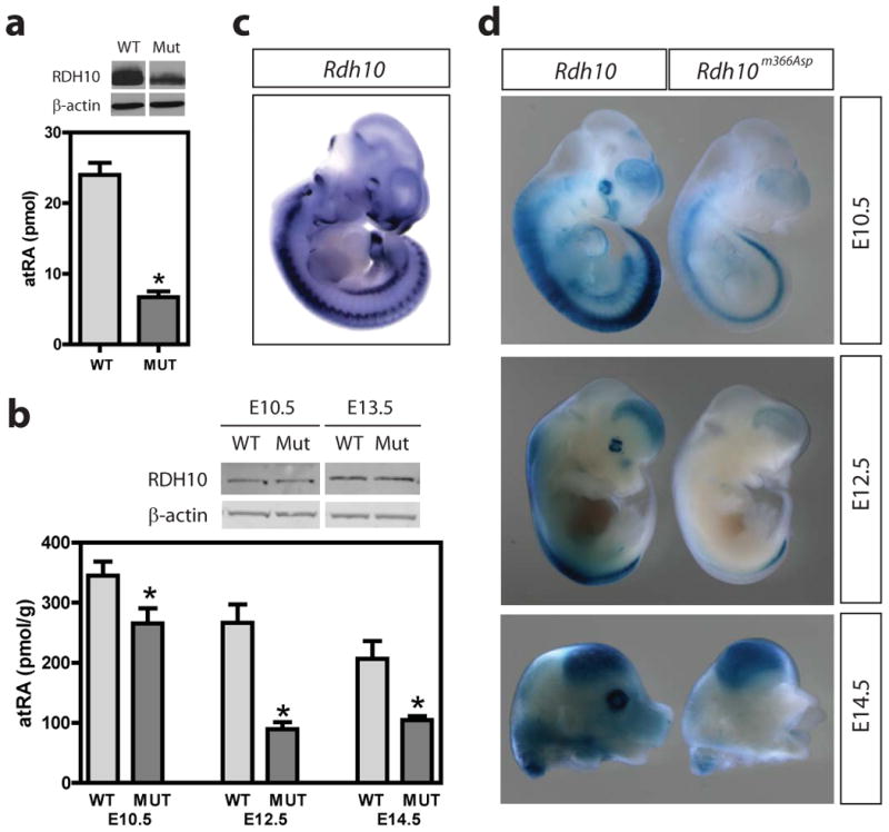
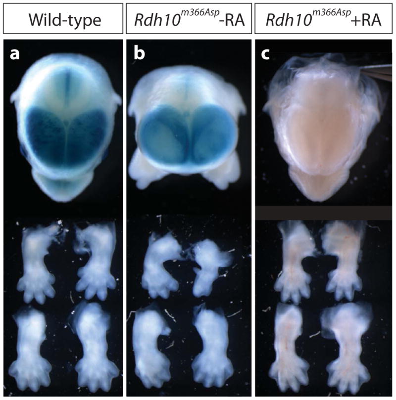
Retinoic acid (RA) signaling is necessary for proper patterning and morphogenesis during embryonic development. Tissue-specific RA signaling requires precise spatial and temporal synthesis of RA from retinal by retinaldehyde dehydrogenases (Raldh) and the conversion of retinol to retinal by retinol dehydrogenases (Rdh) of the short-chain dehydrogenase/reducatase gene family (SDR). The SDR, retinol dehydrogenase 10 (RDH10), is a major contributor to retinal biosynthesis during mid-gestation. We have identified a missense mutation in the Rdh10 gene (Rdh10(m366Asp) ) using an N-ethyl-N-nitrosourea-induced forward genetic screen that result in reduced RA levels and signaling during embryonic development. Rdh10(m366Asp) mutant embryos have unique phenotypes, such as edema, a massive midline facial cleft, and neurogenesis defects in the forebrain, that will allow the identification of novel RA functions.


Figures

Figure 1
Figure 1. Morphological defects in ENU-induced mutant line 366 embryos
Wild-type (WT) (a) and mutant (Mut) (b) embryos at E14.5 expressing a Dlx-LacZ reporter (Zerucha et al., 2000) that was used in the mutagenesis screen (see Zarbalis et al., 2004). (c) Frontal view of a mutant embryo at (E14.5) displaying very prominent midline facial cleft, while the lower jaw forms normally. (d,e) Transverse sections through the developing eyes at E12.5. (f,g) Whole embryos at E16.75. Note the inflated skin in the mutant that is characteristic of edema (g). (h) Left forelimbs from mutant compared to wild-type limb at E14.5 showing example of limb defects, which are variable. (i) Right hindlimb from mutant and wild-type limb showing mild interdigital webbing (asterisk). (j,k) Sagittal section through the brain at E16.5. Asterisks marks the position of the absent olfactory bulbs in mutant (k).
Figure 2
Figure 2. Mutant line 366 carries a mutation in the Rdh10 gene
(a) Base-pair sequence and corresponding amino acid sequence of a region of the Rdh10 gene and RDH10 protein, respectively. Highlighted are the mutated nucleotide and corresponding amino acid. The alanine-to-valine missense mutation is a conservative change, with valine having a pair of methyl groups replacing two hydrogen atoms on the amino acid side chain of the alanine residue. (b) Amino acid #196 of the RDH10 protein is highly conserved amino acid residue. Also highly conserved are the residues surrounding Alanine196. In the segment of RDH10 protein depicted, there is 100% conservation among humans, bovine, rat and mouse (summarized as mammalian). Some divergence begins to appear in amino acids nearby Alanine196 in zebrafish and Xenopus.
Figure 3
Figure 3. The Rdh10m366Asp mutation reduces RA biosynthesis
(a) On top is a representative western blot of N-terminal-FLAG-tagged RDH10 and N-terminal-FLAG-tagged mutant RDH10m366Asp after transfection of CHO cells. Mutant protein levels were consistently 40% less than wild-type, presumably because of either reduced translation and/or stability. At bottom, CHO cells co-transfected with FLAG-tag WT-RDH10 or FLAG-tagged Mut-RDH10m366Asp and RALDH2 biosynthesize atRA. Retinol was incubated with transfected cells for 1 hr (n=3 wells assayed independently). Background atRA values produced by cells transfected with empty vectors and RALDH2 were subtracted to provide net pmol RA. Net pmol RA values for RDH10m366Asp mutant and wild-type RDH10 proteins were normalized to differences in protein levels. A significant difference between wild-type RDH10 and mutant RDH10m366Asp was found, *p = 0.008. Comparable data were reproduced at least three times under identical conditions. (b) On top is a representative western blot of RDH10 protein from whole wild-type or Rdh10m366Asp embryos at E10.5 and E13.5. The RDH10 protein migrates at ∼39kDa, as expected. Unlike our in vitro studies, we did not detect any appreciable difference in RDH10 protein levels from whole embryos as a result of the A196V mutation. At bottom, RA levels in whole wild-type and Rdh10m366Asp embryos at three embryonic stages expressed as pmol/g protein ± SEM. Since morphological defects in Rdh10m366Asp mutants are first apparent after E10.5, as indicated by hypoplasia of the MNPs, we wanted to assay for differences in RA levels just prior to seeing these initial defects. We also examined subsequent stages (E12.5 and E14.5) to determine whether changes in RA levels persist or recover. Number of embryos analyzed at each embryonic age (wild-type/mutant): E10.5, 11/13; E12.5, 6/4; E14.5, 5/5; *p <0.04. (c) Rdh10 expression at E10.5. Note how closely the expression of Rdh10 overlaps with endogenous RA signaling (d) at E10.5. (d) Sagittal view of whole embryos at E10.5 and E12.5, and E14.5 head, stained for β-galactosidase activity after crossing the Rdh10m366Asp mutant to a RA-reporter strain (Rossant et al., 1991). Reduced β-galactosidase staining is observed at all stages. See also Figure 4a,b for anterior view of head at E14.5 after β-galactosidase staining.
Figure 4
Figure 4. Rdh10m366Asp mutant phenotype is rescued by RA supplementation
(a) Top, anterior view of head shows complete formation of facial midline in wild-type mice at E14.5. Bottom, forelimb and hindlimbs. (b) Rdh10m366Asp embryos displaying face, brain and limb phenotypes. (c) Rdh10m366Asp embryos fed a RA supplemented diet. The severe limb and craniofacial defects were rescued to such an extent that they were indistinguishable from wild-type embryos at E14.5. Limb outgrowth and digit patterning were morphologically normal. The midline facial cleft was non-evident as the snout formed normally. In addition, the eyes were the same size as wild-type embryos, and Rdh10m366Asp mutant embryos no longer had a domed forebrain region.

References

    1. Ashique AM, Choe Y, Karlen M, May SR, Phamluong K, Solloway MJ, Ericson J, Peterson AS. The Rfx4 transcription factor modulates Shh signaling by regional control of ciliogenesis. Sci Signal. 2009;2:1–12. - PubMed
    1. Barua AB, Furr HC. Properties of retinoids. Mol Biotechnol. 1998;10:167–182. - PubMed
    1. Belyaeva OV, Johnson MP, Kedishvili NY. Kinetic analysis of human enzyme RDH10 defines the characteristics of a physiologically relevant retinol dehydrogenase. J Biol Chem. 2008;283:20299–308. - PMC - PubMed
    1. Bohnsack BL, Lai L, Dolle P, Hirschi KK. Signaling hierarchy downstream of retinoic acid that independently regulates vascular remodeling and endothelial cell proliferation. Genes Dev. 2004;18:1345–58. - PMC - PubMed
    1. Chatzi C, Brade T, Duester G. Retinoic acid functions as a key GABAergic differentiation signal in the basal ganglia. PLoS Biol. 2011;9:e1000609. - PMC - PubMed

Publication types

LinkOut - more resources